
Page 164 - Read Online
P. 164
Alberts et al. Vessel Plus 2023;7:34 https://dx.doi.org/10.20517/2574-1209.2023.37 Page 3 of 24
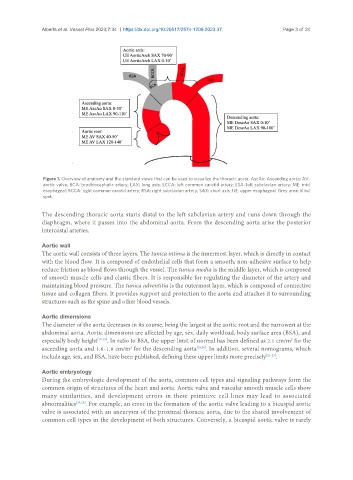
Figure 1. Overview of anatomy and the standard views that can be used to visualize the thoracic aorta. AscAo: Ascending aorta; AV:
aortic valve; BCA: brachiocephalic artery; LAX: long axis; LCCA: left common carotid artery; LSA: left subclavian artery; ME: mid
esophageal; RCCA: right common carotid artery; RSA: right subclavian artery; SAX: short axis, UE: upper esophageal. Grey area: blind
spot.
The descending thoracic aorta starts distal to the left subclavian artery and runs down through the
diaphragm, where it passes into the abdominal aorta. From the descending aorta arise the posterior
intercostal arteries.
Aortic wall
The aortic wall consists of three layers. The tunica intima is the innermost layer, which is directly in contact
with the blood flow. It is composed of endothelial cells that form a smooth, non-adhesive surface to help
reduce friction as blood flows through the vessel. The tunica media is the middle layer, which is composed
of smooth muscle cells and elastic fibers. It is responsible for regulating the diameter of the artery and
maintaining blood pressure. The tunica adventitia is the outermost layer, which is composed of connective
tissue and collagen fibers. It provides support and protection to the aorta and attaches it to surrounding
structures such as the spine and other blood vessels.
Aortic dimensions
The diameter of the aorta decreases in its course, being the largest at the aortic root and the narrowest at the
abdominal aorta. Aortic dimensions are affected by age, sex, daily workload, body surface area (BSA), and
2
especially body height [17-19] . In ratio to BSA, the upper limit of normal has been defined as 2.1 cm/m for the
ascending aorta and 1.6-1.8 cm/m for the descending aorta [18,20] . In addition, several nomograms, which
2
include age, sex, and BSA, have been published, defining these upper limits more precisely [21-23] .
Aortic embryology
During the embryologic development of the aorta, common cell types and signaling pathways form the
common origin of structures of the heart and aorta. Aortic valve and vascular smooth muscle cells show
many similarities, and development errors in these primitive cell lines may lead to associated
abnormalities [24,25] . For example, an error in the formation of the aortic valve leading to a bicuspid aortic
valve is associated with an aneurysm of the proximal thoracic aorta, due to the shared involvement of
common cell types in the development of both structures. Conversely, a bicuspid aortic valve is rarely