


Page 127 - Read Online
P. 127
Cheng et al. Soft Sci. 2026, 6, 36                                                Page 9 of 15


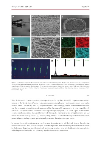
               Figure 3. (A) Photo of conjugate electrospinning preparation of cortisol-sensing yarns; (B) Two rolls of cortisol-sensing yarns prepared
               through continuous conjugate electrospinning technology; (C and D) Surface morphology of cortisol-sensing yarns under low and high
               magnification; (E) Cross-section SEM image and (F) surface elemental mapping of a cortisol-sensing yarn; (G) Photos of dye infiltration
               into a cortisol-sensing yarn within 5 s; (H) WCA of electrospun sensing films within 133 ms. SEM: Scanning electron microscopy; WCA:
               water contact angle.


                                                                                                                                                                 (3)
                                                        P = 2γ cos θ / r
               Here, P denotes the Laplace pressure, corresponding to the capillary force (F ); γ represents the surface
                                                                                   c
               tension of the liquid; θ signifies the instantaneous contact angle; and r indicates the mean pore radius
               between fibers. This capillary force (F ) originates from the surface energy gradient established between sweat
                                              c
               and the nanoscale pores of the sensing cortex, while the permeable nanoporous structure significantly
               enhances this capillary effect, thereby accelerating the capillary dynamics of sweat. Upon initial contact,
               sweat is rapidly drawn into the nanofiber network through the synergistic action of capillary force (F ) and
                                                                                                      c
               omnidirectional wetting forces (F ). Subsequently, sweat is adsorbed onto adjacent fibers and within
                                             w
               interstitial pores, leading to rapid spreading and permeation throughout the yarn cortex.


               In real-world wearable applications, an excellent sweat absorption ability will definitely shorten the collection
               time and incubation time of sweat samples, contributing to real-time monitoring of cortisol concentration
               levels. Besides, the porous nanofiber network morphology creates a large interfacial contact area, conducive
               to binding cortisol molecules and enhancing signal differences and sensitivities.

