


Page 130 - Read Online
P. 130
Page 12 of 15                                                       Cheng et al. Soft Sci. 2026, 6, 36


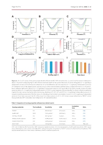
               Figure 5. (A-C) LSV curves of the yarns prepared with different NiHCF-MIP amounts (0.6, 1.2, and 2.4 wt%) tested in blank and 1
               nmol/L~10 μmol/L cortisol. Ag/AgCl as the reference electrode and Pt as the counter electrode; (D) Linear fitting plots (n = 3, signifying 3
               independent sensors); (E) Summary and comparison of current difference (DI) and sensitivity (n = 3, signifying 3 independent sensors);
               (F) Calibration curve of the amperometric response versus cortisol concentration in artificial sweat, ranging from 1 nmol/L to 10 μmol/L.
               Inset: calibration plot with a linear fit (n = 3, signifying 3 independent sensors); (G) Selectivity of the yarns towards cortisol and other
               sweat secretions (n = 3, signifying 3 independent sensors); (H) LSV current response of the sensing fiber to a fixed cortisol concentration
               (1 μmol/L) under various mechanical deformations (e.g., bending at 0°, 30°, 60°, and 90°) (n = 3, signifying 3 independent sensors); (I)
               Long-term stability assessment of the fiber sensor for detecting a fixed cortisol concentration (1 μmol/L) (n = 3, signifying 3 independent
               sensors). Error bars represent the SD of the mean from three sensors in (D-I). LSV: Linear sweep voltammetry; NiHCF: nickel
               hexacyanoferrate; MIP: molecularly imprinted polymer; SD: standard deviation.


               Table 1. Comparison of sensing properties with previous related reports
                                                                                    Incubation
               Sensing materials       Test methods  Sensitivity     LOD                         Refs.
                                                                                    time
               PB-MIP                  CA            38.8 nA·dec -1  0.9 nmol/L     120 s        [15]
               AuNPs-MIP               SWV           2.061 μA·dec -1  200 fmol/L    480 s        [35]
               CNTfiber-PB-MIP         CA            48.7 μA·dec -1  1 pmol/L       120 s        [19]
               MXene/AuNPs-aptamer     DPV           0.389 μA·dec -1  0.276 nmol/L  40 min       [36]
               PB-MIP-PEDOT:PSS        LSV           2.549 μA·dec -1  0.36 nmol/L   150 s        [37]
               NiHCF-MIP               LSV           2.08 μA·dec -1  0.4 nmol/L     120 s        This work

               LOD: Limit of detection; PB: Prussian blue; MIP: molecularly imprinted polymer; CA: chronoamperometry; AuNPs: gold nanoparticles; SWV: square
               wave voltammetry; CNT: carbon nanotube; DPV: differential pulse voltammetry; PEDOT: poly(3,4-ethylenedioxythiophene); PSS:
               poly(styrenesulfonate); LSV: linear sweep voltammetry; NiHCF: nickel hexacyanoferrate.

