
Page 146 - Read Online
P. 146
Page 4 of 15 Ding et al. Soft Sci. 2026, 6, 2
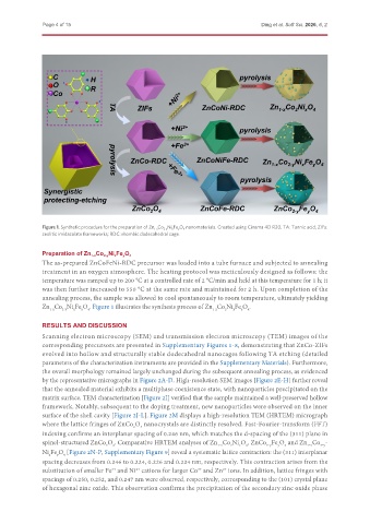
Figure 1. Synthetic procedure for the preparation of Zn 1-x Co 2-y Ni x Fe y O 4 nanomaterials. Created using Cinema 4D R20. TA: Tannic acid; ZIFs:
zeolitic imidazolate frameworks; RDC: rhombic dodecahedral cage.
Preparation of Zn 1-x Co 2-y Ni x Fe y O 4
The as-prepared ZnCoFeNi-RDC precursor was loaded into a tube furnace and subjected to annealing
treatment in an oxygen atmosphere. The heating protocol was meticulously designed as follows: the
temperature was ramped up to 200 °C at a controlled rate of 2 °C/min and held at this temperature for 1 h; it
was then further increased to 550 °C at the same rate and maintained for 2 h. Upon completion of the
annealing process, the sample was allowed to cool spontaneously to room temperature, ultimately yielding
Zn Co Ni Fe O . Figure 1 illustrates the synthesis process of Zn Co Ni Fe O .
4
x
1-x
x
y
4
2
y
2-y
1-x
RESULTS AND DISCUSSION
Scanning electron microscopy (SEM) and transmission electron microscopy (TEM) images of the
corresponding precursors are presented in Supplementary Figures 1-8, demonstrating that ZnCo-ZIFs
evolved into hollow and structurally stable dodecahedral nanocages following TA etching (detailed
parameters of the characterization instruments are provided in the Supplementary Materials). Furthermore,
the overall morphology remained largely unchanged during the subsequent annealing process, as evidenced
by the representative micrographs in Figure 2A-D. High-resolution SEM images [Figure 2E-H] further reveal
that the annealed material exhibits a multiphase coexistence state, with nanoparticles precipitated on the
matrix surface. TEM characterization [Figure 2I] verified that the sample maintained a well-preserved hollow
framework. Notably, subsequent to the doping treatment, new nanoparticles were observed on the inner
surface of the shell cavity [Figure 2J-L]. Figure 2M displays a high-resolution TEM (HRTEM) micrograph
where the lattice fringes of ZnCo O nanocrystals are distinctly resolved. Fast-Fourier-transform (FFT)
2
4
indexing confirms an interplanar spacing of 0.246 nm, which matches the d-spacing of the (311) plane in
spinel-structured ZnCo O . Comparative HRTEM analyses of Zn Co Ni O , ZnCo Fe O and Zn Co -
4
2
1-x
x
4
1‑x
2‑y
y
2
4
2-y
Ni Fe O [Figure 2N-P, Supplementary Figure 9] reveal a systematic lattice contraction: the (311) interplanar
4
y
x
spacing decreases from 0.246 to 0.224, 0.226 and 0.224 nm, respectively. This contraction arises from the
substitution of smaller Fe and Ni cations for larger Co and Zn ions. In addition, lattice fringes with
2+
3+
2+
3+
spacings of 0.250, 0.252, and 0.247 nm were observed, respectively, corresponding to the (101) crystal plane
of hexagonal zinc oxide. This observation confirms the precipitation of the secondary zinc oxide phase