
Page 43 - Read Online
P. 43
Page 14 of 28 Zhang et al. Soft Sci 2024;4:39 https://dx.doi.org/10.20517/ss.2024.34
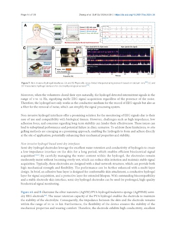
Figure 7. Non-invasive hydrogel interfaces. (A and B) Physically cross-linked interpenetrating network based on calcium ions [92] ; (C and
D) Viscoelastic hydrogel dampers for electrophysiological sensors [79] .
Moreover, when the volunteers closed their eyes naturally, the hydrogel detected intermittent signals in the
range of 9 to 12 Hz, signifying stable EEG signal acquisition regardless of the presence of the noise.
Therefore, the hydrogel not only works as the conductive medium for the record of EEG signals but also as
a filter for the removal of noise, which can simplify the signal processing system.
Non-invasive hydrogel interfaces offer a promising solution for the monitoring of EEG signals due to their
ease of use and compatibility with biological tissues. However, challenges such as high impedance, low
adhesion force, and concerns regarding long-term stability can hinder their effectiveness. These issues can
lead to suboptimal performance and potential failure in clinic scenarios. To address these limitations, in-situ
gelling methods are emerging as a promising approach, enabling the hydrogels to form and adhere directly
at the site of application, potentially enhancing their mechanical properties and stability.
Non-invasive hydrogel-based semi-dry interfaces
Semi-dry hydrogel electrodes leverage the excellent water retention and conductivity of hydrogels to create
a low-impedance interface on the skin for a long period, which enables efficient bioelectrical signal
acquisition [32,93] . By carefully managing the water content within the hydrogel, the electrodes remain
moderately moist without becoming overly wet, which can reduce skin irritation and maintain stable signal
acquisition. Typically, these electrodes are designed with a dual-network structure, which can provide both
high mechanical strength and flexibility. The performance can be further enhanced with a multi-layer
design. In brief, an adhesive base layer is designed for conformable skin attachment, a conductive hydrogel
layer for signal acquisition, and a protective layer for extended lifespan. With outstanding biocompatibility
and a stable electrode-skin interface, semi-dry hydrogel electrodes can be used for prolonged, high-quality
bioelectrical signal monitoring.
Figure 8A and B illustrates the silver nanowire (AgNW)/PVA hydrogel/melamine sponge (AgPHMS) semi-
dry EEG electrode . The water retention capacity of the PVA hydrogel enables the electrode to maintain
[94]
the stability of the electrolyte. Consequently, the impedance between the skin and the electrode remains
within the range of 10 to 15 kΩ. Furthermore, the flexibility of the device ensures the stability of the
mechanical properties and wearing comfort. Therefore, the electrode exhibits high conductivity, excellent