
Page 37 - Read Online
P. 37
Page 8 of 28 Zhang et al. Soft Sci 2024;4:39 https://dx.doi.org/10.20517/ss.2024.34
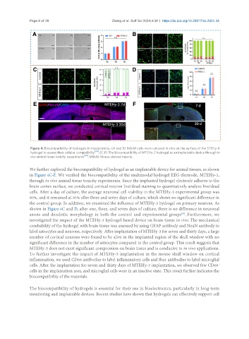
Figure 4. Biocompatibility of hydrogels in bioelectricity. (A and B) MSkM cells were cultured in vitro on the surface of the STEHy-6
hydrogel to assess their cellular compatibility [84] ; (C-F) The biocompatibility of MTEHy-3 hydrogel as an implantable device through in
vivo animal tissue toxicity experiments [85] . MSkM: Mouse skeletal muscle.
We further explored the biocompatibility of hydrogel as an implantable device for animal tissues, as shown
in Figure 4C-F. We verified the biocompatibility of the multimodal hydrogel EEG electrode, MTEHy-3,
through in vivo animal tissue toxicity experiments. Since the implanted hydrogel electrode adheres to the
brain cortex surface, we conducted cortical neuron live/dead staining to quantitatively analyze live/dead
cells. After a day of culture, the average neuronal cell viability in the MTEHy-3 experimental group was
90%, and it remained at 85% after three and seven days of culture, which shows no significant difference in
the control group. In addition, we examined the influence of MTEHy-3 hydrogel on primary neurons. As
shown in Figure 4C and D, after one, three, and seven days of culture, there is no difference in neuronal
axons and dendritic morphology in both the control and experimental groups . Furthermore, we
[85]
investigated the impact of the MTEHy-3 hydrogel-based device on brain tissue in vivo. The mechanical
combability of the hydrogel with brain tissue was assessed by using GFAP antibody and NeuN antibody to
label astrocytes and neurons, respectively. After implantation of MTEHy-3 for seven and thirty days, a large
number of cortical neurons were found to be alive in the implanted region of the skull window with no
significant difference in the number of astrocytes compared to the control group. This result suggests that
MTEHy-3 does not exert significant compression on brain tissue and is conducive to in vivo applications.
To further investigate the impact of MTEHy-3 implantation in the mouse skull window on cortical
inflammation, we used CD68 antibodies to label inflammatory cells and Iba1 antibodies to label microglial
cells. After the implantation for seven and thirty days of MTEHy-3 implantation, we observed few CD68
+
cells in the implantation area, and microglial cells were in an inactive state. This result further indicates the
biocompatibility of the materials.
The biocompatibility of hydrogels is essential for their use in bioelectronics, particularly in long-term
monitoring and implantable devices. Recent studies have shown that hydrogels can effectively support cell