
Page 86 - Read Online
P. 86
Page 10 of 13 Zhao et al. Soft Sci. 2025, 5, 10 https://dx.doi.org/10.20517/ss.2024.61
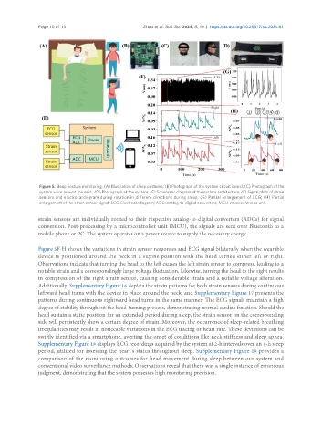
Figure 5. Sleep posture monitoring. (A) Illustration of sleep postures; (B) Photograph of the system circuit board; (C) Photograph of the
system worn around the neck; (D) Photograph of the system; (E) Schematic diagram of the system architecture; (F) Signal plots of strain
sensors and electrocardiogram during rotation in different directions during sleep; (G) Partial enlargement of ECG; (H) Partial
enlargement of the strain sensor signal. ECG: Electrocardiogram; ADC: analog-to-digital converters; MCU: microcontroller unit.
strain sensors are individually routed to their respective analog-to-digital converters (ADCs) for signal
conversion. Post-processing by a microcontroller unit (MCU), the signals are sent over Bluetooth to a
mobile phone or PC. The system operates on a power source to supply the necessary energy.
Figure 5F-H shows the variations in strain sensor responses and ECG signal bilaterally when the wearable
device is positioned around the neck in a supine position with the head turned either left or right.
Observations indicate that turning the head to the left causes the left strain sensor to compress, leading to a
notable strain and a correspondingly large voltage fluctuation. Likewise, turning the head to the right results
in compression of the right strain sensor, causing considerable strain and a notable voltage alteration.
Additionally, Supplementary Figure 16 depicts the strain patterns for both strain sensors during continuous
leftward head turns with the device in place around the neck, and Supplementary Figure 17 presents the
patterns during continuous rightward head turns in the same manner. The ECG signals maintain a high
degree of stability throughout the head-turning process, demonstrating normal cardiac function. Should the
head sustain a static position for an extended period during sleep, the strain sensor on the corresponding
side will persistently show a certain degree of strain. Moreover, the occurrence of sleep-related breathing
irregularities may result in noticeable variations in the ECG tracing or heart rate. These deviations can be
swiftly identified via a smartphone, averting the onset of conditions like neck stiffness and sleep apnea.
Supplementary Figure 18 displays ECG recordings acquired by the system at 2-h intervals over an 8-h sleep
period, utilized for assessing the heart’s status throughout sleep. Supplementary Figure 19 provides a
comparison of the monitoring outcomes for head movement during sleep between our system and
conventional video surveillance methods. Observations reveal that there was a single instance of erroneous
judgment, demonstrating that the system possesses high monitoring precision.