
Page 85 - Read Online
P. 85
Zhao et al. Soft Sci. 2025, 5, 10 https://dx.doi.org/10.20517/ss.2024.61 Page 9 of 13
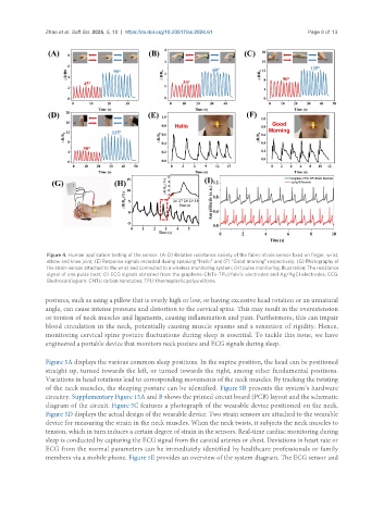
Figure 4. Human application testing of the sensor. (A-D) Relative resistance variety of the fabric strain sensor fixed on finger, wrist,
elbow and knee joint; (E) Response signals recorded during speaking “Hello” and (F) “Good morning” respectively; (G) Photography of
the strain sensor attached to the wrist and connected to a wireless monitoring system; (H) pulse monitoring. Illustration: The resistance
signal of one pulse beat; (I) ECG signals obtained from the graphene-CNTs-TPU/fabric electrodes and Ag/AgCl electrodes. ECG:
Electrocardiogram; CNTs: carbon nanotubes; TPU: thermoplastic polyurethane.
postures, such as using a pillow that is overly high or low, or having excessive head rotation or an unnatural
angle, can cause intense pressure and distortion to the cervical spine. This may result in the overextension
or torsion of neck muscles and ligaments, causing inflammation and pain. Furthermore, this can impair
blood circulation in the neck, potentially causing muscle spasms and a sensation of rigidity. Hence,
monitoring cervical spine posture fluctuations during sleep is essential. To tackle this issue, we have
engineered a portable device that monitors neck posture and ECG signals during sleep.
Figure 5A displays the various common sleep positions. In the supine position, the head can be positioned
straight up, turned towards the left, or turned towards the right, among other fundamental positions.
Variations in head rotations lead to corresponding movements of the neck muscles. By tracking the twisting
of the neck muscles, the sleeping posture can be identified. Figure 5B presents the system’s hardware
circuitry. Supplementary Figure 15A and B shows the printed circuit board (PCB) layout and the schematic
diagram of the circuit. Figure 5C features a photograph of the wearable device positioned on the neck.
Figure 5D displays the actual design of the wearable device. Two strain sensors are attached to the wearable
device for measuring the strain in the neck muscles. When the neck twists, it subjects the neck muscles to
tension, which in turn induces a certain degree of strain in the sensors. Real-time cardiac monitoring during
sleep is conducted by capturing the ECG signal from the carotid arteries or chest. Deviations in heart rate or
ECG from the normal parameters can be immediately identified by healthcare professionals or family
members via a mobile phone. Figure 5E provides an overview of the system diagram. The ECG sensor and