
Page 44 - Read Online
P. 44
Page 26 of 43 Wang et al. Soft Sci 2024;4:41 https://dx.doi.org/10.20517/ss.2024.53
In addition to nanoimprinting, chemical coating is a common method for preparing functional structures
on POF surfaces. Unlike SPR-based fiber optic sensing, functional structures coated on POFs can be utilized
[54]
for a variety of fluorescence-based sensing applications . Recently, Giovannini et al. developed a multi-
sensing platform using three fluorescein isothiocyanate (FITC)-based POF sensors for the efficient
quantification of pH, glucose, and matrix metalloproteinase (MMP) concentrations present in the wound
[206]
exudate [Figure 11C and D]. Moreover, inkjet printing technology is emerging as a promising method
for preparing functional structures on POF surfaces and has been used to fabricate POF-based
photodetector devices. For instance, Kara et al. printed colloidal PbS quantum dots (QDs) on a 1-mm-
diameter POF surface wrapped with graphene, working as infrared phototransistors [Figure 11E]. The
[42]
fabricated device detects photocurrents under laser excitation to monitor scattered light in a multi-mode
POF without interrupting the optical path. In 2023, Wang et al. reported a flexible optoelectronic
multimodal sensor capable of detecting and decoupling proximity, pressure, and temperature signals
[201]
[Figure 11F and G]. This was achieved by integrating an interdigital electrode on the POF surface to detect
capacitance, light intensity, and resistance signals, respectively. This multifunctional and integrated self-
decoupled multimodal POF sensor presents promising opportunities for human-machine-environment
interaction applications. In brief, surface modification-based POF sensors exhibit significant potential for
diverse applications in environmental signal monitoring, particularly due to their flexibility and versatility.
In the future, these sensors are anticipated to play an increasingly vital role in fields such as medical
monitoring, environmental protection, and smart home technologies.
Micro-cylindrical sensors for surgical robots
Surgical robots enhance the precision of surgeons during minimally invasive procedures, addressing
challenges such as low standardization and the high risks associated with manual operations. A robotic
surgical system typically comprises two key components: the robotic arm and the end-effector. The robotic
arm, with its multiple degrees of freedom, is primarily responsible for controlling the manipulation of
surgical instruments . The end-effector, which may include tools such as medical needles, forceps, or
[207]
suturing devices, performs the actual surgical tasks. Precise motion control remains a critical challenge in
current robotic surgical systems, which can be addressed through feedback control by monitoring the
[24]
surgical site . Medical needles, as typical micro-cylindrical surgical instruments, can be equipped with
sensors at the tip to enable real-time monitoring of the surgical environment, ensuring smooth and accurate
operation.
Medical needles are among the most essential and widely used tools in medical procedures. They are
extensively applied in various fields, including blood collection, drug administration, biopsy, and tissue
[208]
ablation . Traditionally, impedance measurements for accurate localization of the needle rely on the
needle body as a conductor , which can constrain electrode design and configuration. This limitation can
[209]
[46]
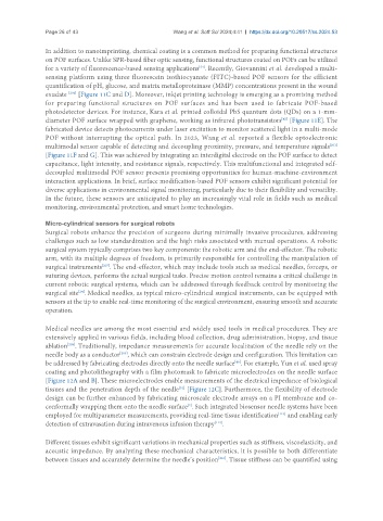
be addressed by fabricating electrodes directly onto the needle surface . For example, Yun et al. used spray
coating and photolithography with a film photomask to fabricate microelectrodes on the needle surface
[Figure 12A and B]. These microelectrodes enable measurements of the electrical impedance of biological
tissues and the penetration depth of the needle [Figure 12C]. Furthermore, the flexibility of electrode
[51]
design can be further enhanced by fabricating microscale electrode arrays on a PI membrane and co-
[2]
conformally wrapping them onto the needle surface . Such integrated biosensor needle systems have been
employed for multiparameter measurements, providing real-time tissue identification and enabling early
[113]
[114]
detection of extravasation during intravenous infusion therapy .
Different tissues exhibit significant variations in mechanical properties such as stiffness, viscoelasticity, and
acoustic impedance. By analyzing these mechanical characteristics, it is possible to both differentiate
between tissues and accurately determine the needle’s position . Tissue stiffness can be quantified using
[210]