
Page 91 - Read Online
P. 91
Lu et al. Soft Sci 2024;4:36 https://dx.doi.org/10.20517/ss.2024.29 Page 11 of 20
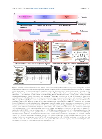
Figure 3. Mechanical evaluation with wide range of measurement depth from superficial surface to deep-tissue sensing. (A) Illustration
of approximate dimensions of the measurement depths targeted to various biological targets via different types of evaluation methods,
involving piezoelectric actuator, mechanical vibration detectors, optical illumination, strain gauge, ultrasonography and MRE, etc; (B)
Photograph of a conformal sensing on a silicone sheet substrate for evaluation of the elastic modulus of epidermis. Reproduced with
[29]
permission . Copyright 2015, Springer Nature; (C) SEM image of the device on an artificial skin sample (upper). A cross-sectional
[29]
illustration of the device (lower). Reproduced with permission . Copyright 2015, Springer Nature; (D) Schematics showing the
[73]
exploded view of a MEMS-based system for the characterization of tissue biomechanics. Reproduced with permission . Copyright
2021, Springer Nature; (E) Dynamic monitoring of human body for modulus sensing. Upper: Photographs of devices on the forearm
[73]
lifting a dumbbell. Lower: sensor Vs as function of time during monitoring. Reproduced with permission . Copyright 2021, Springer
Nature; (F) Exploded-view schematic illustration of the wearable imager for cardiac function assessment, with key elements in terms of
[74]
1-3 piezoelectric composite, electrodes and substrate. Reproduced with permission . Copyright 2023, Springer Nature; (G) M-mode
[74]
images (upper) extracted from wearable imager and corresponding ECG signals (lower). Reproduced with permission . Copyright
2023, Springer Nature; (H) UoC designs for whole body sensing. Photos, SEM images, and schematic diagrams of an UoC design (left)
equipped with 8960 MEMS transducers (upper right) on a CMOS chip, each enabled to send and receive ultrasound signals according
to control/processing circuitry (lower right). Reproduced with permission [65] . Copyright 2021, American Chemical Society; (I) Example
ultrasound imaging by the UoC, abdominal: vascular flow in kidney. Reproduced with permission [65] . Copyright 2021, American Chemical
Society. MRE: Magnetic resonance elastography; SEM: scanning electron microscope; MEMS: micro-electromechanical system; Vs:
voltages; AMVL: anterior mitral valve leaflet; LVPW: left ventricular posterior wall; LVIDd: left ventricular internal diameter end
diastole; LVIDs: left ventricular internal diameter end systole; IVS: interventricular septum; ECG: electrocardiogram; UoC:
ultrasound-on-chip; CMOS: complementary metal-oxide-semiconductor.