
Page 95 - Read Online
P. 95
Lu et al. Soft Sci 2024;4:36 https://dx.doi.org/10.20517/ss.2024.29 Page 15 of 20
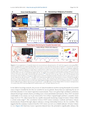
Figure 4. Clinical applications of wearable devices for tissue mechanics characterization. (A) Left: User request access using FENG-
based microphone for voice code recognition. Right: Sound wave of voice code. Reproduced with permission [41] . Copyright 2017, Springer
Nature; (B) Left: Photographs of a piezoelectric device mounted on the forearm to map the lesion regions of dermatologic malignancy.
Right: Spatial mapping of modulus corresponding to measurements on the forearm. Reproduced with permission [29] . Copyright 2015,
Springer Nature; (C) Left: Illustration of the ultrasound sensor mounted on the neck for measurement of pulse waveform from CA,
external jugular vein (ext. JV) and internal jugular vein (int. JV). Right: pulse waveforms of ECG and BP at the artery to assess vascular
stiffness accessibly and reliably. Reproduced with permission [20] . Copyright 2018, Springer Nature; (D) Left: Photographs of an
autonomous sensor holding on the mouse. Right: Closely correlation between resistance readouts from the sensor and the tumor
volume. Reproduced with permission [76] . Copyright 2022, American Association for the Advancement of Science; (E) Left: Photograph of
the device (chest and limb units) on a neonate in a NICU isolette, with exemplary data displaying on a user interface. Middle:
Representative data collected by chest and limb units. Right: Rotational angles between the device and reference frames for a neonate in
NICU in different body positions. Reproduced with permission [77] . Copyright 2020, Springer Nature. FENG: Ferroelectric nanogenerator;
CA: carotid artery; ECG: electrocardiogram; BP: blood pressure; NICU: neonatal intensive care unit.
In the field of oncology research, the process of clinical translation involves testing thousands of potential
cancer drugs to identify the few that are suitable for use in patients. Researchers are exploring the use of
tools that automate in vivo tumor regression measurements. These tools gather high-resolution continuous
datasets from larger animal cohorts, providing more robust and detailed information. Here, FAST mounted
comfortably on the mouse can be pre-stretched to 50% of their original length and have a sensitive strain
range of 25% to 75%, which means they are capable of precisely measuring changes in tumor volume within
a certain strain range . The correlation between FAST sensor resistance readouts and tumor volume
[76]
measurements obtained through calipers indicates a strong relationship between the two variables. This
suggests that the resistance readouts from the FAST sensor can serve as a reliable indicator of tumor volume
in vivo [Figure 4D]. Real-time tumor regression datasets have the potential to significantly enhance the