
Page 87 - Read Online
P. 87
Lu et al. Soft Sci 2024;4:36 https://dx.doi.org/10.20517/ss.2024.29 Page 7 of 20
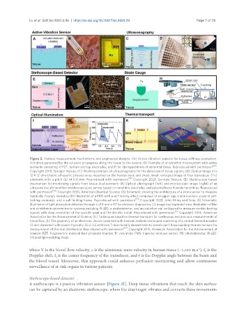
Figure 2. Various measurement mechanisms and engineered designs. (A) Active vibration sensors for tissue stiffness evaluation.
Vibration generated by the actuator propagates along the tissue to the sensor; (B) Example of an ultrathin microsystem with active
[28]
elements consisting of PZT, bottom and top electrodes, and PI for distinguishment of abnormal tissue. Reproduced with permission .
Copyright 2018, Springer Nature; (C) Working principle of ultrasonography for the detection of tissue signals; (D) Optical image of a
12 × 12 stretchable ultrasonic phased array mounted on the human neck and chest. Inset: enlarged image of four transducer (Tx)
elements with a pitch (λ) of 0.8 mm. Reproduced with permission [52] . Copyright 2021, Springer Nature; (E) Stethoscope-based
mechanism for monitoring signals from tissue displacement; (F) Optical photograph (left) and microscopic image (right) of an
ultrasensitive all-nanofiber mechanoacoustic sensor based on nanofibre electrodes and polyvinylidene fluoride nanofibres. Reproduced
with permission [54] . Copyright 2020, American Chemical Society; (G) Schematic showing the architecture of a strain sensor to measure
haptically Young’s modulus; (H) Illustration of a FMS with a self-locking effect composed of an upper cap, a strain sensor, a pair of self-
locking elements, and a self-locking frame. Reproduced with permission [55] . Copyright 2021, John Wiley and Sons; (I) Schematic
illustration of light absorption detected through a LED and a PD for photonic diagnostics; (J) Image and exploded-view illustration of thin
and stretchable optoelectronic systems including IR LED, a photodetector, and an inductive coil configured to measure cardiac beating
signals with clear revelation of the systolic peak and the dicrotic notch. Reproduced with permission [31] . Copyright 2016, American
Association for the Advancement of Science; (K) Techniques based on thermal transport for continuous and precious measurements of
blood flow; (L) The geometry of an electronic device combined with thermal analysis techniques consisting of a central thermal actuator
-2
(3 mm diameter) with power (typically 25 or 3.5 mW·mm ) directionally downstream to vessels and 14 surrounding thermal sensors for
measurement of thermal distribution. Reproduced with permission [63] . Copyright 2015, American Association for the Advancement of
Science. PZT: Piezoelectric material lead zirconate titanate; PI: polyimide; FMS: fingertip modulus sensor; PD: photodetector; IR LED:
infrared light-emitting diode.
where V is the blood flow velocity, c is the ultrasonic wave velocity in human tissue (~1,540 m·s ), f is the
-1
d
Doppler shift, f is the center frequency of the transducer, and θ is the Doppler angle between the beam and
0
the blood vessel. Moreover, this approach could enhance perfusion monitoring and allow continuous
surveillance of at-risk organs in various patients.
Stethoscope-based detector
A stethoscope is a passive vibration sensor [Figure 2E]. Deep tissue vibrations that reach the skin surface
can be captured by an electronic stethoscope, where the diaphragm vibrates and converts these movements