
Page 107 - Read Online
P. 107
Kim et al. Soft Sci 2023;3:16 https://dx.doi.org/10.20517/ss.2023.07 Page 19 of 30
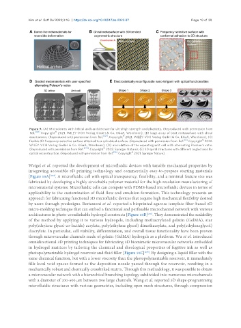
Figure 9. (A) Microdomes with helical arch architecture for ultrahigh strength and plasticity. (Reproduced with permission from
©
Ref. [226] . Copyright 2021. WILEY-VCH Verlag GmbH & Co. KGaA, Weinheim); (B) large array of bent metasurface with chiral
©
enantiomers. (Reproduced with permission from Ref. [227] . Copyright 2021. WILEY-VCH Verlag GmbH & Co. KGaA, Weinheim); (C)
©
flexible 3D frequency-selective surface attached to a cylindrical surface. (Reproduced with permission from Ref. [57] . Copyright 2022.
WILEY-VCH Verlag GmbH & Co. KGaA, Weinheim); (D) microlattice of the repeating unit cell with alternating Poisson’s ratio.
[228] ©
(Reproduced with permission from Ref. . Copyright 2022. Springer Nature); (E) 3D spiral structures with different angled arcs for
[56] ©
optical reconstruction. (Reproduced with permission from Ref. . Copyright 2021. Springer Nature).
Weigel et al. reported the development of microfluidic devices with tunable mechanical properties by
integrating accessible 3D printing technology and commercially easy-to-prepare starting materials
[234]
[Figure 10A] . A microfluidic cell with optical transparency, flexibility, and a minimal feature size was
fabricated by developing a highly stretchable polymer material for the high-resolution manufacturing of
micromaterial systems. Microfluidic cells can compete with PDMS-based microfluidic devices in terms of
applicability to the customization of fluid flow and emulsion formation. This technology presents an
approach for fabricating functional 3D microfluidic devices that require high mechanical flexibility desired
by users through predesigns. Bertassoni et al. reported a bioprinted agarose template fiber-based 3D
micro-molding technique that can embed a functional and perfusable microchannel network with various
architectures in photo-crosslinkable hydrogel constructs [Figure 10B] . They demonstrated the scalability
[235]
of the method by applying it to various hydrogels, including methacrylated gelatin (GelMA), star
poly(ethylene glycol-co-lactide) acrylate, poly(ethylene glycol) dimethacrylate, and poly(ethyleneglycol)
diacrylate. In particular, cell viability, differentiation, and overall tissue functionality have been proven
through microvascular channels made of gelatin (GelMA) hydrogels as a platform. Wu et al. introduced
omnidirectional 3D printing techniques for fabricating 3D biomimetic microvascular networks embedded
in hydrogel matrices by tailoring the chemical and rheological properties of fugitive ink as well as
photopolymerizable hydrogel reservoir and fluid filler [Figure 10C] . By designing a liquid filler with the
[236]
same chemical function, but with a lower viscosity than the photopolymerizable reservoir, it immediately
fills local void spaces formed as the deposition nozzle passed through the reservoir, resulting in a
mechanically robust and chemically crosslinked matrix. Through this methodology, it was possible to obtain
a microvascular network with a hierarchical branching topology subdivided into numerous microchannels
with a diameter of 200-600 μm between two large channels. Wang et al. reported 3D shape-programming
microfluidic structures with various geometries, including open mesh structures, through compression