
Page 235 - Read Online
P. 235
Siegal et al. Plast Aesthet Res 2019;6:25 I http://dx.doi.org/10.20517/2347-9264.2019.35 Page 11 of 20
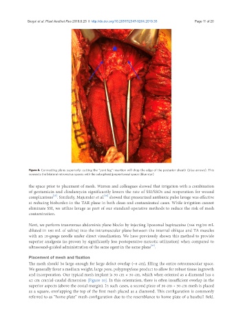
Figure 8. Connecting plans superiorly: cutting the “pant leg” insertion will drop the edge of the posterior sheath (blue arrows). This
connects the bilateral retrorectus spaces with the subxiphoid/preperitoneal space (blue star)
the space prior to placement of mesh. Warren and colleagues showed that irrigation with a combination
of gentamicin and clindamycin significantly lowers the rate of SSI/SSOs and reoperation for wound
[25]
[26]
complications . Similarly, Majumder et al. showed that pressurized antibiotic pulse lavage was effective
at reducing bioburden in the TAR plane in both clean and contaminated cases. While irrigation cannot
eliminate SSI, we utilize lavage as part of our standard operative methods to reduce the risk of mesh
contamination.
Next, we perform transversus abdominis plane blocks by injecting liposomal bupivacaine (266 mg/20 mL
diluted in 180 mL of saline) into the intramuscular plane between the internal oblique and TA muscles
with an 18-gauge needle under direct visualization. We have previously shown this method to provide
superior analgesia (as proven by significantly less postoperative narcotic utilization) when compared to
ultrasound-guided administration of the same agent in the same plane .
[27]
Placement of mesh and fixation
The mesh should be large enough for large defect overlap (~8 cm), filling the entire retromuscular space.
We generally favor a medium weight, large pore, polypropylene product to allow for robust tissue ingrowth
and incorporation. Our typical mesh implant is 30 cm × 30 cm, which when oriented as a diamond has a
42 cm cranial-caudal dimension [Figure 10]. In this orientation, there is often insufficient overlap in the
superior aspects (above the costal margin). In such cases, a second piece of 30 cm × 30 cm mesh is placed
as a square, overlapping the top of the first mesh placed as a diamond. This configuration is commonly
referred to as “home plate” mesh configuration due to the resemblance to home plate of a baseball field.