
Page 232 - Read Online
P. 232
Page 8 of 20 Siegal et al. Plast Aesthet Res 2019;6:25 I http://dx.doi.org/10.20517/2347-9264.2019.35
Figure 5. Lateral dissection: the lateral dissection can proceed as far back as the psoas muscle in the retroperitoneum
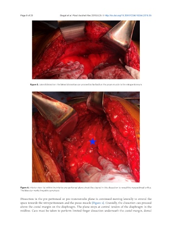
Figure 6. Inferior view: fat within the inferior pre-peritoneal plane should be cleared in this dissection to reveal the myopectineal orifice.
The blue star marks the pubic symphysis
Dissection in the pre-peritoneal or pre-transversalis plane is continued moving laterally to extend the
space towards the retroperitoneum and the psoas muscle [Figure 5]. Cranially, the dissection can proceed
above the costal margin on the diaphragm. The plane stops at central tendon of the diaphragm in the
midline. Care must be taken to perform limited finger dissection underneath the costal margin, dorsal