
Page 229 - Read Online
P. 229
Siegal et al. Plast Aesthet Res 2019;6:25 I http://dx.doi.org/10.20517/2347-9264.2019.35 Page 5 of 20
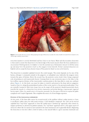
Figure 1. Entering the retrorectus space: After palpating the edge of the rectus muscle, the rectus sheath is incised 5-10 mm lateral to the
medial edge revealing muscle fibers
retraction (tension) is evenly distributed and less likely to tear fascia. Blunt and electrocautery dissection
is then used to extend the dissection to the lateral edge of the recuts muscle where the linea semilunaris is
encountered. Working lateral, it is helpful to have the assistant use a Richardson retractor to lift the rectus
up and away from the posterior sheath as the surgeon moves laterally. Kittner (peanut) dissectors are
helpful tools to sweep the loose alveolar tissue off the posterior sheath as one works laterally.
The dissection is extended cephalad towards the costal margin. The extent depends on the size of the
hernia, although it commonly extends to the epigastric or subxiphoid area. Inferiorly, the surgeon works
towards the space of Retzius. Below the arcuate line, the posterior rectus sheath thins (being composed
only of peritoneum and transversalis fascia). Crossing from the one retrorectus space to the other in the
low midline requires division of the transversalis insertion points to the linea alba to create one confluent
plane [Figure 2]. At this level, care must be taken to identify and preserve the deep inferior epigastric
vessels as they run along the posterolateral surface of the rectus muscle in the pretransversalis plane. They
are typically invested in fibro fatty tissue that can be swept off the posterior sheath/transversalis fascia
towards the muscle in a dissection that mimics maneuvers performed during a laparoscopic inguinal
hernia repair. Often, the caudal extent of dissection proceeds into the space of Retzius to expose the pubis
symphysis and Cooper’s ligaments. This completes the extent of a Rives-Stoppa exposure.
Division of the transversus abdominis
At this point, if the linea alba cannot be reconstructed in the midline without undue tension or there
is insufficient sublay space for wide mesh overlap, a TAR should be completed. The TAR can be started
cephalad first (“top-down” approach) or from the caudal aspect (“bottom-up” approach), often chosen by
surgeon preference or dictated by patient anatomy. The “top-down” approach starts in the upper aspect of
dissection where the TA muscle is more medial to the linea semilunaris and is generally thicker. Beginning
the dissection at this level offers a level of safety, as the muscle belly is a good anatomic landmark and