
Page 231 - Read Online
P. 231
Siegal et al. Plast Aesthet Res 2019;6:25 I http://dx.doi.org/10.20517/2347-9264.2019.35 Page 7 of 20
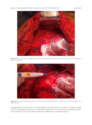
Figure 3. Accessing the transversus abdominis: the posterior lamella of the internal oblique is scored (blue arrow) to reveal the underlying
transversus abdominis
Figure 4. Releasing the transversus abdominis: a right angle clamp gentle reveals the muscle fibers that are then divided with
electrocautery
propagating tears once they start can be challenging to fix). Either plane can be used, and surgeons should
become comfortable learning how to “plane-hop” between these two as needed on a patient-by-patient
basis and based on the need to address focal areas of difficulty in any individual patient.