
Page 327 - Read Online
P. 327
Liu et al. Adipogenesis of obital fat stem cells
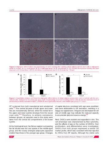
A B
C D
Figure 3: Adipogenic differentiation was assessed by oil red O staining after culturing orbital adipose-derived stem cells in adipogenic
media for 14 days. (A, C): young group; (B, D): middle-aged group; (A, B): non-induced; (C, D): induced. More positive staining cells were
observed in (A, C) than those in (B, D), and almost no staining was seen in (A, B) (scale bar: 100 µm)
A Induced B Induced
Non-induced Non-induced
Figure 4: Quantitative analysis confirmed the adipogenic differentiation of orbital adipose-derived stem cells in contrast with the non-
induced cells. Compared to the young age group, both the percentage of oil red O staining-positive cells (A) and the expression level of
peroxisome proliferator-activated receptor γ mRNA (B) were significantly reduced in the middle-aged group (*P < 0.001)
OF is derived from both mesodermal and ectodermal of septal structure correlated with age were predicted,
cells. [13] The central fat pads of both upper and lower and were attributable to OF herniation, resulting in a
lids are mesodermally derived, while the medial fat in baggy-eye appearance. Irregular and decreased fat cell
the upper and lower eyelids develops from the neural size at middle age may indicate the reduced capacity
crest cells. [4,5] Therefore, to achieve consistency to accumulate lipid and reserve energy. [14]
between groups, all samples used in this study were
taken from the central fat compartments of the lower Next, OASCs were isolated and expanded in vitro. The
eyelids. cell phenotype was characterized by flow cytometry,
and the effects of age on the number of OASCs’, their
At the histological level, the fibrous septum surrounding proliferation, and differentiation were investigated.
the fat lobules was thin and sparse in the middle-aged OASCs from both groups expressed a similar CD
group, and the loosely arranged adipocytes appeared marker profile, which was consistent with that reported
smaller than those in the younger age group. Changes for ASCs from SF depots. Although the viable cells
326 Plastic and Aesthetic Research ¦ Volume 3 ¦ October 25, 2016