
Page 318 - Read Online
P. 318
Agrawal et al. Total septal reconstruction using costal cartilage
Surgical technique
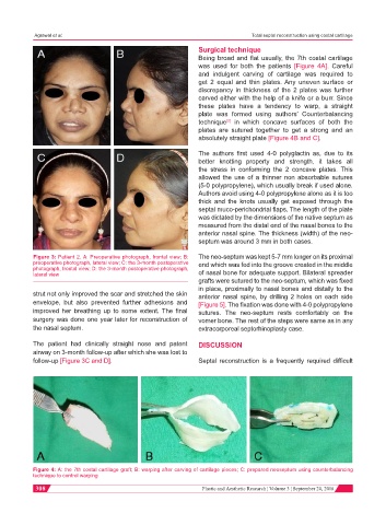
Being broad and flat usually, the 7th costal cartilage
was used for both the patients [Figure 4A]. Careful
and indulgent carving of cartilage was required to
get 2 equal and thin plates. Any uneven surface or
discrepancy in thickness of the 2 plates was further
carved either with the help of a knife or a burr. Since
these plates have a tendency to warp, a straight
plate was formed using authors’ Counterbalancing
technique in which concave surfaces of both the
[2]
plates are sutured together to get a strong and an
absolutely straight plate [Figure 4B and C].
The authors first used 4-0 polyglactin as, due to its
better knotting property and strength, it takes all
the stress in conforming the 2 concave plates. This
allowed the use of a thinner non absorbable sutures
(5-0 polypropylene), which usually break if used alone.
Authors avoid using 4-0 polypropylene alone as it is too
thick and the knots usually get exposed through the
septal muco-perichondrial flaps. The length of the plate
was dictated by the dimensions of the native septum as
measured from the distal end of the nasal bones to the
anterior nasal spine. The thickness (width) of the neo-
septum was around 3 mm in both cases.
Figure 3: Patient 2. A: Preoperative photograph, frontal view; B: The neo-septum was kept 5-7 mm longer on its proximal
preoperative photograph, lateral view; C: the 3-month postoperative end which was fed into the groove created in the middle
photograph, frontal view; D: the 3-month postoperative photograph,
lateral view of nasal bone for adequate support. Bilateral spreader
grafts were sutured to the neo-septum, which was fixed
in place, proximally to nasal bones and distally to the
strut not only improved the scar and stretched the skin anterior nasal spine, by drilling 2 holes on each side
envelope, but also prevented further adhesions and [Figure 5]. The fixation was done with 4-0 polypropylene
improved her breathing up to some extent. The final sutures. The neo-septum rests comfortably on the
surgery was done one year later for reconstruction of vomer bone. The rest of the steps were same as in any
the nasal septum. extracorporeal septorhinoplasty case.
The patient had clinically straight nose and patent DISCUSSION
airway on 3-month follow-up after which she was lost to
follow-up [Figure 3C and D]. Septal reconstruction is a frequently required difficult
Figure 4: A: the 7th costal cartilage graft; B: warping after carving of cartilage pieces; C: prepared neoseptum using counterbalancing
technique to control warping
308 Plastic and Aesthetic Research ¦ Volume 3 ¦ September 20, 2016