
Page 315 - Read Online
P. 315
Sharma et al. Hard palate cysts
undiagnosed for a long period because of their
asymptomatic background. CT and MRI modalities have
4 brought revolution in diagnosing these entities while
1 performed for some other reasons. These should be
2
classified in their proper category before treating them.
Financial support and sponsorship
3 None.
Conflicts of interest
There are no conflicts of interest.
Patient consent
The consent of the patient was taken before subjecting
the patient for investigation.
Ethics approval
The approval for publishing this case and paper had
been obtained from the institute.
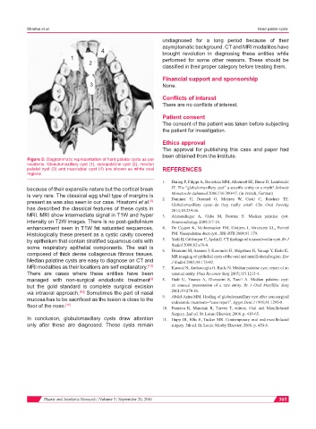
Figure 5: Diagrammatic representation of hard palatal cysts as per
locations. Globulomaxillary cyst (1), nasopalatine cyst (2), median
palatal cyst (3) and nasolabial cyst (4) are shown as white oval REFERENCES
regions
1. Haring P, Filippi A, Bornstein MM, Altermatt HJ, Buser D, Lambrecht
because of their expansile nature but the cortical break JT. The “globulomaxillary cyst” a specific entity or a myth? Schweiz
is very rare. The classical egg shell type of margins is 2. Monatsschr Zahnmed 2006;116:380-97. (in French, German)
Dammer U, Driemel O, Mohren W, Giedl C, Reichert TE.
present as was also seen in our case. Hisatomi et al. Globulomaxillary cysts--do they really exist? Clin Oral Investig
[6]
has described the classical features of these cysts in 2014;18:239-46.
MRI. MRI show intermediate signal in T1W and hyper 3. Alimendinger A, Gabe M, Destian S. Median palatine cyst.
intensity on T2W images. There is no post-gadolinium Neuroradiology 2009;3:7-10.
enhancement seen in T1W fat saturated sequences. 4. De Cuyper K, Vanhoenacker FM, Hintjens J, Verstraete KL, Parizel
Histologically these present as a cystic cavity covered PM. Nasopalatine duct cyst. JBR-BTR 2008;91:179.
by epithelium that contain stratified squamous cells with 5. Yerli H, Cabbarpur C, Aydin E. CT findings of a nasoalveolar cyst. Br J
some respiratory epithelial components. The wall is 6. Radiol 2009;82:e76-8.
Hisatomi M, Asaumi J, Konouchi H, Shigehara H, Yanagi Y, Kishi K.
composed of thick dense collagenous fibrous tissues. MR imaging of epithelial cysts of the oral and maxillofacial region. Eur
Median palatine cysts are easy to diagnose on CT and J Radiol 2003;48:178-82.
MRI modalities as their locations are self explanatory. [7,8] 7. Karacal N, Ambarcoglu O, Kutlu N. Median palatine cyst: report of an
There are cases where these entities have been unusual entity. Plast Reconstr Surg 2005;115:1213-4.
managed with non-surgical endodontic treatment 8. Hadi U, Younes A, Ghosseini S, Tawil A. Median palatine cyst:
[9]
but the gold standard is complete surgical excision an unusual presentation of a rare entity. Br J Oral Maxillfac Surg
via intraoral approach. Sometimes the part of nasal 2001;39:278-81.
[10]
mucosa has to be sacrificed as the lesion is close to the 9. Abdel-Azim MM. Healing of globulomaxillary cyst after non-surgical
endodontic treatment--“case report”. Egypt Dent J 1995;41:1295-8.
floor of the nose. [11] 10. Fonseca R, Marciani R, Turvey T, editors. Oral and Maxillofacial
Surgery. 2nd ed. St. Loius: Elsevier; 2008. p. 418-65.
In conclusion, globulomaxillary cysts draw attention 11. Hupp JR, Ellis E, Tucker MR. Contemporary oral and maxillofacial
only after these are diagnosed. These cysts remain surgery. 5th ed. St. Louis: Mosby Elsevier; 2008. p. 450-6.
Plastic and Aesthetic Research ¦ Volume 3 ¦ September 20, 2016 305