
Page 317 - Read Online
P. 317
Agrawal et al. Total septal reconstruction using costal cartilage
INTRODUCTION
Rhinoplasty and septal reconstruction often require the
use of cartilage grafts. Occasionally, the unsuspecting
rhinoplasty surgeon may hitherto stumble upon such
noses which have paucity of native cartilaginous and
bony septum. In post-septoplasty severely deviated
nose and severely deformed post traumatic nose, the
native septal cartilage and bony septum may be thin,
fragmented or inadequate. The reconstruction of a
complete septum with costal cartilage may restore a
strong support in such situations. Here we describe
two such cases.
CASE REPORT
Case 1
A 26-year-old male had a history of injury to the nose by
cricket ball 5 years ago. His nose had gradually deviated
and owing to severe breathing problems, he underwent
septoplasty 2 years ago. There was considerable relief
in breathing after the surgery but the nose remained
crooked. He presented to us with a crooked nose for
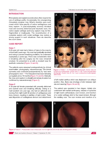
aesthetic correction [Figure 1A and B].
The patients were assessed postoperatively by clinical
examination, photography, nasoendoscopy. The dorsal Figure 1: Patient 1. A: Preoperative photograph, frontal view; B:
symmetry was confirmed postoperatively by “text neck preoperative photograph, lateral view; C: the 1-year postoperative
photographic view”. The first patient has been following photograph, frontal view; D: the 1-year postoperative photograph,
[1]
lateral view
up regularly since 18 months [Figure 1C and D]. Clinically
the nose and septum is straight and airway is patent as
confirmed by nasoendoscopy [Figure 2]. of left medial canthus which was displaced in an oblique
position. Also, there was shortage of skin between right
Case 2 ala and nasal tip [Figure 3A and B].
A 35-year-old female presented with severely deformed
and scarred nose and breathing difficulty. Owing to a The patient was operated in two stages. Initially she
road accident one year ago, she had an adherent scar underwent left medial canthopexy, release of synechia
running from right nostril till junction of cartilaginous and of right nostril, adhesiolysis, scar revision, and insertion
bony dorsum, resulting in partition of right nostril. There of a costal cartilage strut to the nasal dorsum, through
was disjunction of cartilaginous dorsum and detachment the existing scar. The scar revision and insertion of a
Figure 2: Patient 1. Postoperative naso-endoscopic view on right (A) and left (B) side showing patent airway
Plastic and Aesthetic Research ¦ Volume 3 ¦ September 20, 2016 307