
Page 314 - Read Online
P. 314
Sharma et al. Hard palate cysts
A B C
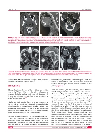
Figure 3: Non contrast computerized tomography of facial and neck region in 3D reformatted bone window. (A) Axial section shows
expanded radiolucent lesion with “egg shell” appearance at the base of nasal region predominantly on right side (vertical white arrow); (B)
sagittal section shows the superior extent of lesion from hard palate (white star); (C) coronal section delineate the lesion separate from the
maxillary sinuses but obscuring the nasal passages predominantly on right side (white star)
A B C
Figure 4: Plain magnetic resonance imaging face and neck region. (A) T2W image shows the well contained hyperintense cystic lesion on
right side of the hard palate (vertical arrow); (B) T2W sagittal image shows hyperintense well demarcated lesion on the superior part of the
hard palate (white star) on right side; (C) same lesion in T2WI coronal section with its superior extent (vertical arrow)
enucleation of the cyst as this being the most preferred fusion of upper jaw bones. Non-odontogenic cysts can
[3]
method of treatment of these entities. further be differentiated on the basis of their anatomical
location. All the cysts around incisive canal fall in this
DISCUSSION group [Figure 5].
Hard palate forms the floor of the nostrils and roof of the Globulomaxillary cysts arise at the junction between
oral cavity. This is thicker in front and thin in its posterior maxilla and premaxilla. There are three main subtypes
aspect. Globulomaxillary cysts are still disputable in of non-odontogenic fissural cysts described as
[4]
their origin but majority of studies have shown these as nasoalveolar, nasopalatine and median palatal.
not of embryonic origin. [2] These are usually discovered during routine clinical
or radiographic examinations. The average duration
Oral origin cysts can be placed in to two categories as of these cysts vary from one week to two years. The
follow: (1) non-odontogenic (fissural) category includes occlusal images are the first to lead in radiological
globulomaxillary, nasopalatine, median palatal and investigations. In plain X-ray this may present as
nasolabial (nasoalveolar) cysts; and (2) odontogenic radiolucent region. These are usually asymptomatic and
category includes dentigerous cysts, primordial cysts, also found as incidental findings in CT examinations.
odontogenic keratocysts and residual cysts. Their pathogenesis though controversial but non
disintegrated epithelium in the fissural sites remains the
Globulomaxillary cysts fall in non-odontogenic category. most accepted hypothesis. These are usually painless
These can be distinguished based on the origin of the and rarely get infected and that is the reason for their
epithelial rests. Odontogenic cysts arise from tooth delayed diagnosis. These are oval or round and
[5]
developing epithelium contrary to non odontogenic hypodense on CT examination and do not enhance in
which arise from the trapped epithelium because of the post contrast studies. Bone resorption is often present
304 Plastic and Aesthetic Research ¦ Volume 3 ¦ September 20, 2016