
Page 303 - Read Online
P. 303
Hwang et al. Medially based de-epithelialized flap
c’
sn sbal al
sbal A
ac
Figure 3: Four anthropometric distances were measured: nostril
floor width (Sbal-Sn), alar distance (Sn-Al), alar curvature distance
(Sn-Ac), and nostril length (Sbal-C’). al: alare; ac: alar curvature
point; sn: subnasale; sbal: subalare; c’: highest point of the columella
columellar base, fresh epithelium was shaved on the
medial side of the incision line. The widened scar on
the upper lip was excised. The raw (denuded and de-
epithelialized) tip of the columellar base was pulled
under the medial tip of the alar base flap and sutured
tightly. The nasal base was then narrowed and the
nostril sill was augmented [Figure 2]. B
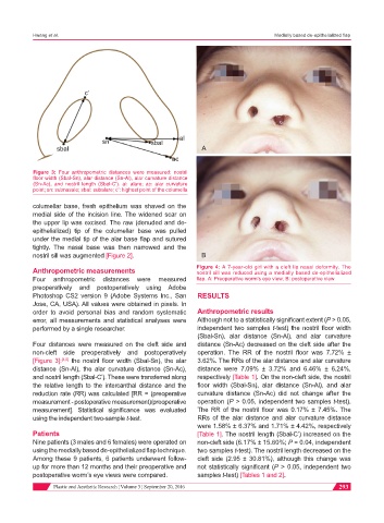
Anthropometric measurements Figure 4: A 7-year-old girl with a cleft lip nasal deformity. The
nostril sill was reduced using a medially based de-epithelialized
Four anthropometric distances were measured flap. A: Preoperative worm’s eye view; B: postoperative view
preoperatively and postoperatively using Adobe
Photoshop CS2 version 9 (Adobe Systems Inc., San RESULTS
Jose, CA, USA). All values were obtained in pixels. In
order to avoid personal bias and random systematic Anthropometric results
error, all measurements and statistical analyses were Although not to a statistically significant extent (P > 0.05,
performed by a single researcher. independent two samples t-test) the nostril floor width
(Sbal-Sn), alar distance (Sn-Al), and alar curvature
Four distances were measured on the cleft side and distance (Sn-Ac) decreased on the cleft side after the
non-cleft side preoperatively and postoperatively operation. The RR of the nostril floor was 7.72% ±
[Figure 3]: [4,5] the nostril floor width (Sbal-Sn), the alar 3.62%. The RRs of the alar distance and alar curvature
distance (Sn-Al), the alar curvature distance (Sn-Ac), distance were 7.09% ± 3.72% and 6.46% ± 6.24%,
and nostril length (Sbal-C’). These were transferred along respectively [Table 1]. On the non-cleft side, the nostril
the relative length to the intercanthal distance and the floor width (Sbal-Sn), alar distance (Sn-Al), and alar
reduction rate (RR) was calculated [RR = (preoperative curvature distance (Sn-Ac) did not change after the
measurement - postoperative measurement)/preoperative operation (P > 0.05, independent two samples t-test).
measurement]. Statistical significance was evaluated The RR of the nostril floor was 0.17% ± 7.45%. The
using the independent two-sample t-test. RRs of the alar distance and alar curvature distance
were 1.58% ± 6.37% and 1.71% ± 4.42%, respectively
Patients [Table 1]. The nostril length (Sbal-C’) increased on the
Nine patients (3 males and 6 females) were operated on non-cleft side (6.17% ± 15.60%; P = 0.04, independent
using the medially based de-epithelialized flap technique. two samples t-test). The nostril length decreased on the
Among these 9 patients, 6 patients underwent follow- cleft side (2.95 ± 30.81%), although this change was
up for more than 12 months and their preoperative and not statistically significant (P > 0.05, independent two
postoperative worm’s eye views were compared. samples t-test) [Tables 1 and 2].
Plastic and Aesthetic Research ¦ Volume 3 ¦ September 20, 2016 293