
Page 21 - Read Online
P. 21
Peritoneal Membrane, W.L. Gore and Associates. Inc.),
between the dura and the soft tissues, especially at the site
of the temporal muscle. [16]
Extradural and extracranial pooling of fluid and subdural
hematoma are less frequent events in cranioplasties. The
former can usually be resolved by prompting parenchymal
re‑expansion (if viable) or by increasing the number of
dural suspension points and maintaining subcutaneous
drainage for a longer period of time. Adhesion of the
scalp to the cranial implant can be promoted by anchoring
the latter to the galea fascia using sutures.
The soft tissues overlying the cranioplasty implant can
also be subject to ischemia, necrosis, and/or decubitus,
and it is thus vital that cutaneous trophism and irrigation
is carefully evaluated in the presurgical phase. Moreover, a Figure 3: “Italic S” technique. If the cranioplasty involves the use of
surgical approach should be planned taking into account more pieces faced between them, the contact surfaces must not be
not only aesthetic concerns (such as avoiding the incision linear. This prevents slips and dislocations
encroaching below the hairline and using the Simpson
technique) but also seeking to avoid damage to the main
arterial trunks and temporal muscle. [13,17] In difficult cases
featuring a paucity of viable soft tissue, cranioplasty
implant fitting could necessitate the use of cutaneous
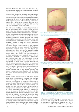
expanders. Another useful surgical aid for improving
cutaneous trophism is dermal matrix (INTEGRA Dermal
Regeneration Template Single Layer film) [Figure 5].
[18]
Such matrices promote mesenchymal histoinduction and
histoconduction, serving to guide the formation of normal
dermal tissue. The collagen and glucosaminoglycans
of these matrices provide structural support for the
infiltrating fibroblasts, macrophages, lymphocytes,
and capillaries that form the neurovascular network.
In covering the implant, these networks favor the
development of better blood irrigation, important not
only for cutaneous tropism but also for the invasion of Figure 4: In the pterional area, the anchorage of the temporalis muscle
should not be done on the cranioplasty, but must override it, with
the porous HA of the cranial implant by the organic bone traction to the sagittal line
matrix, promoting osteoconduction and osteointegration
of the prosthesis. The scalp is not only necessary for
implant coverage but it also supplies nutrients and
immune system components. Together with the dura, it
also aids in the osteomimesis process of the cranioplasty
implant.
Indeed, another possible cause of HA cranial implant
failure is lack of osteomimesis. If there is poor contiguity
between the implant and the skull margin, osteoblast
migration is compromised. To avoid this and to ensure the
accurate design of the implant (which must fit perfectly
along the entire border of its cranial housing), the
surgeon must take certain factors into account during the
surgery itself. In particular, the skull defect borders must
be cleared completely of any scarring or inflammatory
matrix, the dura on the border of the internal plate
must be delaminated, and the craniectomy border drilled Figure 5: The trophism of the skin overlying the cranioplasty is
delicately. In addition, no material should be placed important for the osteomimesis and for the prevention of infections.
The trophism of the skin may improve by using dermal matrix placed
between the bone and implant, with the exception of HA between cranioplasty and subcutaneous tissue
granules or calcium phosphate paste [Figure 6]. Indeed, it
has been demonstrated that more osteointegration occurs In fact, the threshold for damage to osteocytes is as low
on a rough surface. A prime concern of the surgeon, as 47 °C. That being said, the limited clinical success
[20]
[19]
however, should be that the continuum is controlled and of osteomimesis could also be explained by a lack of
that the tissue exposed to drilling is adequately cooled. vascularization, which is affected by the tropism of the
10 Plast Aesthet Res || Vol 2 || Issue 1 || Jan 15, 2015