
Page 20 - Read Online
P. 20
implants are indicated. The incidence of infection was first 3 months or after 6 months. The time between
1.77%, a finding comparable to that reported for titanium 3 and 6 months is associated with the highest risk of
implants (1.18%) and slightly better than that for PMMA complications, both infectious and otherwise.
prostheses (5.48%). [3‑7] Of the infections, 73% occurred Another complication arising from cranioplasty is the
during the 1st year after fitting, confirming that infection
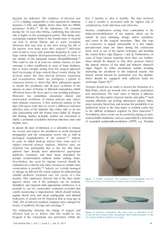
risk is higher in the postsurgical period. That being said, dislocation/mobilization of the implant, which can be
cranioplasty implants fitted in the frontal sinus or mastoid caused by poor planning, design and/or validation,
can lead to airway fistulas and to acute secondary and errors in the surgical procedure. Thus, this type
infections that may arise at any time during the life of of occurrence is largely preventable if a few simple
the patient, even many years after surgery. [8,9] Infections precautionary steps are taken during the craniotomy
were found to occur with particular frequency in cases of itself, such as use of the jigsaw technique and beveling
large cranial implants (frontoparietotemporal), or those in the cranial defect edge [Figures 1 and 2]. Furthermore, in
the vicinity of the paranasal sinuses (frontal/bifrontal). cases of large lacunas requiring more than one implant,
[10]
This could be due to at least two distinct factors: (1) skin these should be shaped so that their juncture mimics
coverage is often insufficient in cases of large implants, the natural sutures of the skull and features slanted‑S
due to tissue atrophy arising from the surgical approach edges [Figure 3]. Other precautions include avoiding
itself (sectioning of large arterial blood vessels during the anchoring the prosthesis to the temporal muscle; this
incision) and/or the time interval between craniotomy muscle should instead be positioned over the implant,
and reconstruction, which can predispose a patient to which should be equipped with sufficient holes for
cutaneous lesions or ulcers that allow pathogenic agents anchorage [Figure 4]. [13]
to invade the prosthesis; and (2) poor occlusion of the Attempts should also be made to prevent the formation of a
sinuses, in cases of frontal or bifrontal cranioplasty, which fluid fistula, which can severely slow or impede cicatrisation
effectively leaves the door open to any invading pathogen. and osteomimesis. The main cause of fistulas is adhesion
Moreover, the sometimes precarious clinical and between the dura mater, temporal muscle, and galea. Such
[14]
neurological conditions of trauma patients may reduce scarring adhesions can prolong subsequent surgery times,
their immune responses. A first statistical analysis of the cause excessive blood loss, and increase the probability of an
data (Chi‑square test) did not reveal a difference between inadvertent lesion to the dura mater or cerebral cortex due
infection rates of HA implants that either take or do not to the difficult techniques required for their dissection.
[15]
take relationship with the frontal sinus [Table 3]. Despite Nevertheless, these events can be averted by placing an inert,
this finding, further in‑depth, studies are warranted to nonresorbable membrane, such as a super‑thin (0.1 mm) sheet
clarify a potential correlation between infection rates and
implant sites. of expanded polytetrafluoroethylene (ePTFE; e.g. Preclude
In almost all cases of infection, it is advisable to cleanse
the wound and remove the prosthesis to avoid intradural
propagation and the consequent severe risk as well as
prolonged hospitalization of the patient. [8,11] Indeed,
in cases in which back‑up devices have been used to
replace removed primary implants, infection rates are
relatively low, presumably due to the fact that these
patients have already been administered appropriate
antibiotic treatment and have been scheduled for
prompt re‑intervention without undue waiting times.
Nevertheless, the need for implant removal should be
evaluated on a case‑by‑case basis, because in certain cases
conservation is possible. Indeed, we recently managed
[12]
to salvage an infected HA cranial implant by administering
suitable antibiotic treatment over the course of a few
months. This experience showed that if the dura mater Figure 1: “Puzzle” technique. The perimeters of cranioplasty must be
appears intact, and if the pathogen can be isolated, characterized by extroflexions to prevent slips and dislocations
identified, and targeted with appropriate antibiotics, it is
possible to opt for conservative treatment provided that
careful monitoring is implemented, which should include
regular blood tests and serial scintigraphy with labeled
leukocytes. It should not be forgotten that as long ago as
1948, 25% of infected synthetic implants were salvaged by
means of antibiotic therapy and curettage. [8]
The relationship between the timing of surgery and
infection lead us to believe that this would be less Figure 2: Forehead cranioplasty. The edges have an inclination of 45°
to prevent the sinking of the cranioplasty. Male forehead profile on the
frequent if the cranioplasty was performed within the left; female forehead profile on the right
Plast Aesthet Res || Vol 2 || Issue 1 || Jan 15, 2015 9