
Page 19 - Read Online
P. 19
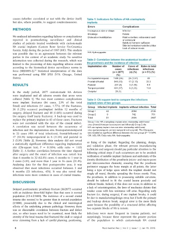
causes  (whether correlated or not with the device itself)   Table 1: Indicators for failure of HA cranioplasty
          but also, where possible, to suggest countermeasures.  implants
                                                               Errors                  Complications
          METHODS                                              Incongruous size or shape  Infection
                                                               Breakage                Fistula
          We analyzed information regarding failures or complications   Dislocation/mobilization  Fluid collection: extracranial and/
          reported  in  postmarketing  surveillance  and  clinical                     or extradural
          studies of patients treated worldwide with custom‑made                       Subdural hematic suffusion
          HA  cranial implants  (Custom Bone Service Fin‑Ceramica                      Skin ischemia/necrosis/decubitus
                                                                                       Lack of osteomimesis
          Faenza, Italy) during the period of 1997‑2013. This analysis
          was possible due to an agreement between the relevant   HA: Hydroxyapatite
          parties in the context of an academic study. No sensitive
          information was collected during the research, which was   Table 2: Correlation between the anatomical location of
          limited to the processing of data regarding adverse events   the prosthesis and the incidence of infection
                                                                                           Cases of  Rates in total
          according  to  the  biomedical  device  surveillance  norms  in   Implantation site   Number of   infections   infections
                                                                                  cases
                         [2]
          force  (MEDDEV‑2).  Statistical interpretation of the data            (n = 2877)   (n) (%)  (n = 51) (%)
          was  performed  using  IBM  SPSS  (V19;  Chicago, United                 (%)
          States).                                             Fontoparietotemporal  1588 (55)  25 (1.57)  49
                                                               Frontal-bifrontal  548 (19)  17 (3.10)   33.3
          RESULTS                                              Parietal           231 (8)   3 (1.30)     5.9
                                                               Temporoparietal   491 (17)   6 (1,22)    11.8
          In the  study period, 2877 custom‑made HA devices    Occipital          29 (1)       -         -
          were  implanted  and all adverse  events  that  arose  were
          collated  [Table  1].  The  two  most  common  complications   Table 3: Chi-square test to compare the infections
          were implant fractures  (84  cases, 2.9% of the total   implant rates of two groups
          fitted) and infections  (51  cases, 1.77%).  Of the fractures,   Group  Infected implants Implants without infection Total
          36  (1.25%)  occurred postimplant  (within 12  months of   Group 1  42              2094         2136
          surgery, delayed fracture) and 48 (1.66%) occurred during   Group 2  9              742           751
          the surgery itself (early fractures). A back‑up was used to   Total  51             2836         2887
          replace the primary implant in 43 of these cases. Fractures
          were not correlated with the size  of the cranial defect.   Group 1: the HA cranioplasty implant takes relationship with frontal
                                                               sinus (frontal, frontoparietotemporal and bifrontal); Group 2: the
          A  correlation  was  noted  between  the  occurrence  of   HA cranioplasty implant does not take relationship with frontal
          infection and the implantation site: frontoparietotemporal   sinus (parietotemporal, parietal, temporal and occipital).  The Chi‑square
          in  25 cases  (49% of total infections),  frontal‑bifrontal in   test revealed no significant difference between the two groups (P = 0.1694;
          17 (33.3%) temporoparietal in 6  (11.8%)  and parietal in   OR 1.65). OR: Odds ratio; HA: Hydroxyapatite
          3 (5.9%)  [Table 2]. However, data analysis did not reveal   or  fixing  of  the  implant.  Regarding  the  planning,  design,
          a statistically significant difference regarding implantation   and validation phase, the relevant persons  (manufacturing
          site  (Chi‑square  test; P  =  0.1694; odds ratio  =  1.65)
          [Table 3]. A further correlation between the time elapsed   technician and surgeon) should pay particular attention to the
          after  surgery  and the  onset  of infection  was noted: less   following critical steps if such occurrences are to be avoided:
          than 6 months in 32 (62.8%) cases, 6 months to 1 year in   verification of suitable implant thickness and uniformity of the
          3 cases (5.8%), and more than 1 year in 16 cases (31.4%).   density distribution of the prosthesis (micro‑ and macro‑pores
          Analyzing data for the first postoperative year, it was   and  interconnection  channels),  ensuring  that  the  prosthesis
          observed that most infections occurred between  3 and   perimeter engages the bone margin at all points, the latter
          6  months (23  infections, 45%).  It was also  noted that   being a type of ledge upon which the implant should rest
          infections were more common in cases of cranial trauma.  snugly all round, thereby spreading the forces evenly. Thus,
                                                              the prosthesis, in addition to possessing suitable curvature,
          DISCUSSION                                          should be tailored to fit the cranial lacuna precisely and
                                                              without breaks. Indeed, if this does not occur, in addition to
                                                              a lack of osteointegration, the laws of mechanics dictate that
          Delayed posttraumatic prosthesis fracture (36/2877) occurred   weaker areas with less resistance  will arise. Regarding early
          with an incidence three‑fold higher than that seen in normal   fracture  (i.e.  during surgery), if one implant breaks, it could
          population  (3.5‑4.5/1000). The incidence of a second cranial   be due to manufacturing/design error, but if both the primary
          trauma also seemed to be greater than in normal population   and  back‑up  devices  break,  surgical  error  is the  more  likely
          (2/1000), presumably due to the clinical and neurological   cause because the possibility of a structural defect affecting
          effects  of  the  underlying  primary  pathology.  However, there   two separate blocks of HA is remote.
          was no discernable correlation between fracture and defect
          size, so other issues need to be examined, most likely the   Infections were more frequent in trauma patients, not
          severity of the head trauma that fractured the skull or surgical   surprisingly, because these  represent the greater portion
          error stemming from a lack of careful planning, positioning,   of the population in which custom‑made HA cranial

            8                                                            Plast Aesthet Res || Vol 2 || Issue 1 || Jan 15, 2015