
Page 16 - Read Online
P. 16
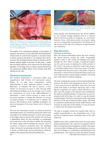
Figure 11: (a) Trachea wrapped with lateral thoracic artery flap, located
                                                              within the lateral thoraco-abdominal area; (b) two constructs can be
                                                              tunneled to two separate lateral neck incisions

                                                              Strap muscles and investing fascia are closed. Rabbits
                                                              do not tolerate foreign material such as a Penrose
                                                              drain to prevent possible air trapping. As a preventive
                                                              measure, loose, interrupted sutures are placed, taking
            Figure 10: Wrapping of an orthotopic tracheal segment within the
            lateral thoracic artery flap. The native trachea is not manipulated until   care not to impair flap vascularization. The distal part of
            complete ingrowth of the flap, i.e. after two weeks. After prefabrication,   the incision is left open for a distance of approximately
            the tracheal segment can be safely manipulated on its pedicle, without   one centimeter.
            the risk of devascularization

            The quality of the anastomosis depends on the degree of   Flap dissection
            exposure, the presence of a bloodless field, and well-prepared   Heterotopic prelamination
            tracheal ends. To control both segments, a retraction suture   After the thoraco-abdominal region has been shaved,
            is placed proximal and distal to the segment that will be   the  lateral  thoracic  vessels  are  easily  recognizable
            removed. The circularly-detached trachea is elevated and the   [Figures 6 and 7]. The vessels are palpable and visible
            desired tracheal length is procured. At this point, a sterile   through the skin. When in doubt, a handheld Doppler
            tube providing isoflurane can be placed into the distal native   can be used to assist in marking the visible portion of
            segment. A suctioning device is used to prevent blood from   the vessels. The point at which the vessels dive deeper
            the submucosal capillary plexus of the tracheal ends from   to reach the axillary artery is the pivot point of the flap.
            leaking into the tube.                            The length from the pivot point to the native trachea is
                                                              measured to ensure that the fabricated flap reaches the
            Tracheal anastomosis                              neck without tension. Extra length is added to the distal
            The tracheal anastomosis is performed under loop   part of the flap for tracheal wrapping.
            magnification with Prolène 6-0 interrupted sutures   The skin overlying the vessels is incised and undermined
            [Figure  5]. As  with  every  anastomosis,  careful   between the dermis and subcutaneous fascia. Once the
            approximation of both segments without overlap is   correct plane has been identified, dissection proceeds
            important.  To  minimize  the  risk  of  stenosis,  as  few   easily from distal to proximal. Operating clips or fine
            sutures as necessary are used to close the gap while   bipolar coagulation are used to divide branches to the
            preventing the leakage of air. On average, 6 to 7 points   skin. Once the length of the flap has been established,
            per anastomosis are used. If the caliber-difference is   the pedicle is divided distally. It is important to preserve
            substantial, a short vertical incision is made in the   enough width of the flap to be able to wrap the desired
            narrowest segment to enlarge the diameter. To prevent   tracheal length. The flap is elevated from the underlying
            secondary healing, it is important not to damage the   muscles via the intervening bloodless plane. Elevation
            mucosa.  Microsurgical  tissue  handling  techniques  are   is continued towards the pivot point while leaving a cuff
            used and grasping of the inner lumen with a forceps is   of tissue on each side of the pedicle. It is not necessary
            avoided.                                          to skeletonize the pedicle proximally.

            The first two interrupted sutures are placed posteriorly.   The flap is  wrapped around the tracheal tube  with its
            Full-thickness bites are avoided by passing the suture from   pedicle perpendicular to  the  longitudinal  axis  of  the
            externally to the submucosal space. As such, the integrity   trachea to facilitate future orthotopic inset of the tube
            of the fragile mucosa is preserved, and the risk of stenosis   [Figure 8].
            is  diminished.  Knots  are  tied  externally,  as  intraluminal
            knots and suture ends will obstruct airflow. By suctioning   Rabbits tend to bite wounds in the trunk. To avoid trauma
            the lumen, again without harming the mucosa, stasis of   during prelamination, the construct is tunneled with its
            secretions or blood is prevented. Next, the remaining   flap to a lateral neck incision [Figures 6 and 8]. This region
            sutures are placed, progressing anteriorly. Once the tube   is not accessible to biting and has an abundance of excess
            is closed, a tracheoscopy can be performed to check the   skin. The technique is a combination of prelamination and
            quality of the anastomosis from the luminal side.  prefabrication.  The trajectory  from  the  lateral thoracic
            228                                                                 Plast Aesthet Res || Volume 3 || July 7, 2016