
Page 15 - Read Online
P. 15
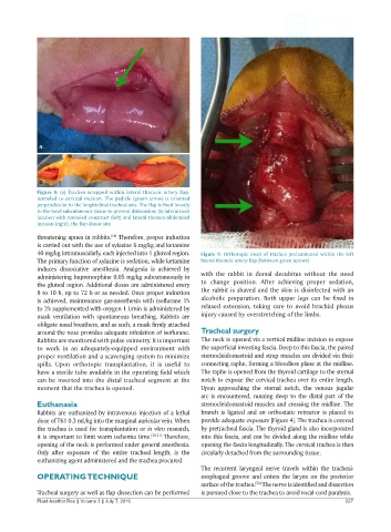
Figure 8: (a) Trachea wrapped within lateral thoracic artery flap,
            tunneled to cervical incision. The pedicle (green arrow) is oriented
            perpendicular to the longitudinal tracheal axis. The flap is fixed loosely
            to the local subcutaneous tissue to prevent dislocation; (b) lateral neck
            incision with tunneled construct (left) and lateral thoraco-abdominal
            incision (right), the flap donor site
            threatening apnea in rabbits.  Therefore, proper induction
                                   [19]
            is carried out with the use of xylazine 6 mg/kg and ketamine
            40 mg/kg intramuscularly, each injected into 1 gluteal region.   Figure 9. Orthotopic inset of trachea prelaminated within the left
            The primary function of xylazine is sedation, while ketamine   lateral thoracic artery flap (between green arrows)
            induces  dissociative  anesthesia. Analgesia is achieved by
            administering buprenorphine 0.05 mg/kg subcutaneously in   with  the  rabbit  in  dorsal decubitus without  the  need
            the gluteal region. Additional doses are administered every   to change position. After achieving  proper sedation,
            8 to 10 h, up to 72 h or as needed. Once proper induction   the rabbit is shaved and the skin is disinfected with an
            is achieved, maintenance gas-anesthesia with isoflurane 1%   alcoholic  preparation. Both upper legs can be fixed in
            to 2% supplemented with oxygen 1 L/min is administered by   relaxed extension, taking care to avoid brachial plexus
            mask ventilation with spontaneous breathing. Rabbits are   injury caused by overstretching of the limbs.
            obligate nasal breathers, and as such, a mask firmly attached
            around the nose provides adequate inhalation of isoflurane.   Tracheal surgery
            Rabbits are monitored with pulse oximetry. It is important   The neck is opened via a vertical midline incision to expose
            to  work  in  an  adequately-equipped environment  with   the superficial investing fascia. Deep to this fascia, the paired
            proper ventilation and a scavenging system to minimize   sternocleidomastoid and strap muscles are divided via their
            spills. Upon orthotopic transplantation,  it  is  useful to   connecting raphe, forming a bloodless plane at the midline.
            have a sterile tube available in the operating field which   The raphe is opened from the thyroid cartilage to the sternal
            can be inserted into the distal tracheal segment at the   notch to expose the cervical trachea over its entire length.
            moment that the trachea is opened.                Upon approaching the sternal notch, the venous jugular
                                                              arc is encountered, running deep to the distal part of the
            Euthanasia                                        sternocleidomastoid muscles and crossing the midline. The
            Rabbits are euthanized by intravenous injection of a lethal   branch is ligated and an orthostatic retractor is placed to
            dose of T61 0.3 mL/kg into the marginal auricular vein. When   provide adequate exposure [Figure 4]. The trachea is covered
            the trachea is used for transplantation or in vitro research,   by pretracheal fascia. The thyroid gland is also incorporated
            it is important to limit warm ischemia time. [20,21]  Therefore,   into this fascia, and can be divided along the midline while
            opening of the neck is performed under general anesthesia.   opening the fascia longitudinally. The cervical trachea is then
            Only after exposure of the entire tracheal length, is the   circularly detached from the surrounding tissue.
            euthanizing agent administered and the trachea procured.
                                                              The recurrent laryngeal nerve travels within the tracheal-
            OPERATING TECHNIQUE                               esophageal groove and enters the larynx on the posterior
                                                              surface of the trachea.  The nerve is identified and dissection
                                                                               [22]
            Tracheal surgery as well as flap dissection can be performed   is pursued close to the trachea to avoid vocal-cord paralysis.
            Plast Aesthet Res || Volume 3 || July 7, 2016                                                 227