
Page 13 - Read Online
P. 13
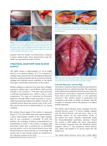
Figure 4: (a) Peroperative view on the pretracheal fascia with the
                                                              thyroid gland (green arrow), after dividing the strap muscles; (b) image
                                                              of posterior oesophageal branches (yellow arrow) after opening the
                                                              pretracheal fascia


            Figure 3: (a) Dissection of the lateral thoracic artery flap. The flap
            is isolated for 2 weeks within the donor area; (b) patent artery and
            vein after 2 weeks of flap isolation; (c) injection of silicone dye into
            the lateral thoracic artery; (d) patent artery with blue silicone dye up
            to distal from the bifurcation and anastomosis with the superficial
            inferior epigastric artery (green arrow)
            Together with the relative cost-effectiveness compared
            to larger animal models, these characteristics make the
            rabbit our experimental model of choice.

            TRACHEAL ANATOMY AND BLOOD
            SUPPLY

            The rabbit trachea is approximately 6.5 cm in length
            and 0.5 cm in diameter [Figure 1].  It is composed of
                                         [10]
            cartilage rings connected by intercartilaginous ligaments
            anteriorly, and the trachealis muscle posteriorly. Both the   Figure 5: Peroperative image of autologous rabbit tracheal transplantation.
                                                              Each anastomosis was closed with 6 points, using interrupted Prolène 6-0
            cartilage and trachealis muscle contribute to the typical   sutures with external knots. The result shows no caliber mismatch
            semi-rigid, semi-flexible characteristic of the trachea.
                                                              Lateral thoracic artery flap
            Hyaline cartilage is composed of an outer layer of highly-  The authors’ workhorse flap for tracheal revascularization
            organized  collagen  type  I  and  II  fibrils,  which  provide   in humans is the free radial forearm flap. The counterpart
            strength to the construct.  This outer layer surrounds   in the rabbit is the pedicled lateral thoracic artery flap. The
                                  [11]
            a hydrated proteoglycan-core, which is able to resist   lateral thoracic artery flap is composed of subcutaneous
            compression. The rings are connected by fibroelastic   fascia with  a thin  muscular layer,  i.e.  the  panniculus
                                                                      [14]
            ligaments, through which vessels penetrate to create a   carnosus.   This  striated  muscle  is  particularly well
            richly-anastomosing submucosal capillary network. The   developed in the trunk of rabbits. In humans, with the
            trachealis muscle shapes the posterior part of the circle.   exception of remnants such as the platysma, it is almost
            This smooth muscle alters luminal diameter, e.g. during   entirely regressed.
            coughing.
                                                              The rabbit’s lateral thoracic  artery originates from the
                                                              external thoracic artery.  It travels caudally  together
                                                                                   [14]
            The intrinsic blood  supply of the trachea is segmental   with  the lateral thoracic vein  and nerve,  deep to the
            [Figure 2].  As such, unlike other solid organ transplants,   pectoralis  muscle. On the latero-inferior border of
                    [12]
            a direct microvascular transfer of the  organ together   the muscle, the artery continues superficially within
            with its own vasculature is unfeasible. To solve this   the thoracoabdominal panniculus carnosus. Around
            problem, the technique of indirect revascularization or   the second to third nipple, the artery anastomoses
            prelamination is used. [5,13]  The trachea is wrapped with   with  secondary  branches  from  the  superficial  inferior
            heterotopic tissue that is perfused by an identifiable   epigastric artery. [15,16]  One prominent lateral thoracic
            vascular pedicle, such as the lateral thoracic fascia with   vein, which runs parallel  to its artery, drains into the
            the lateral thoracic artery and vein. In a second stage, the   axillary vein.
            trachea and its new vascular pedicle are transferred to
            the orthotopic position in the neck.              The human  lateral thoracic artery  has a small caliber
            Plast Aesthet Res || Volume 3 || July 7, 2016                                                 225