
Page 14 - Read Online
P. 14
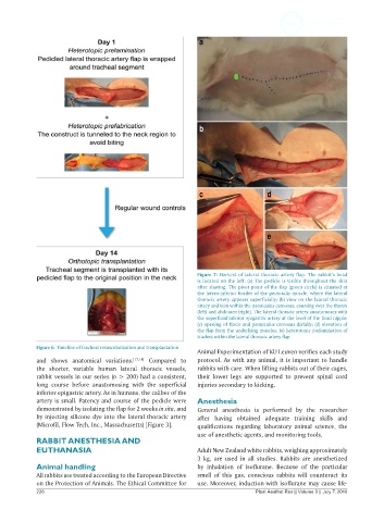
Figure 7: Harvest of lateral thoracic artery flap. The rabbit’s head
                                                              is located on the left. (a) The pedicle is visible throughout the skin
                                                              after shaving. The pivot point of the flap (green circle) is situated at
                                                              the latero-inferior border of the pectoralis muscle, where the lateral
                                                              thoracic artery appears superficially; (b) view on the lateral thoracic
                                                              artery and vein within the panniculus carnosus, coursing over the thorax
                                                              (left) and abdomen (right). The lateral thoracic artery anastomoses with
                                                              the superficial inferior epigastric artery at the level of the third nipple;
                                                              (c) opening of fascia and panniculus carnosus distally; (d) elevation of
                                                              the flap from the underlying muscles; (e) heterotopic prelamination of
                                                              trachea within the lateral thoracic artery flap

            Figure 6: Timeline of tracheal revascularization and transplantation
                                                              Animal Experimentation of KU Leuven verifies each study
            and shows anatomical variations. [17,18]  Compared to   protocol. As with any animal, it is important to handle
            the shorter, variable human lateral thoracic  vessels,   rabbits with care. When lifting rabbits out of their cages,
            rabbit vessels in our series (n > 200) had a consistent,   their  lower legs are supported to prevent spinal cord
            long course before anastomosing  with  the superficial   injuries secondary to kicking.
            inferior epigastric artery. As in humans, the caliber of the
            artery is small. Patency and course of the pedicle were   Anesthesia
            demonstrated by isolating the flap for 2 weeks in situ, and   General anesthesia is performed by the researcher
            by injecting silicone dye into the lateral thoracic artery   after having obtained adequate training skills and
            (Microfil, Flow Tech, Inc., Massachusetts) [Figure 3].  qualifications regarding laboratory animal science, the
                                                              use of anesthetic agents, and monitoring tools.
            RABBIT ANESTHESIA AND
            EUTHANASIA                                        Adult New Zealand white rabbits, weighing approximately
                                                              3 kg, are used in all  studies. Rabbits are anesthetized
            Animal handling                                   by  inhalation  of isoflurane.  Because of the  particular
            All rabbits are treated according to the European Directive   smell of this  gas, conscious rabbits  will counteract its
            on the Protection of Animals. The Ethical Committee for   use. Moreover, induction with isoflurane may cause life-
            226                                                                 Plast Aesthet Res || Volume 3 || July 7, 2016