
Page 83 - Read Online
P. 83
histopathologic margins exclusively analyzing all close and intrinsic factors, such as tissue composition, have been
positive margins. In this study, the mean discrepancy for studied as responsible of different shrinkage percentages
buccal mucosa was 47.6%, 33.3% for the tongue, 9.5% for the at different locations in the given specimen. It has been
mandibular alveolus, and 4.8% for both, retromolar trigone observed that intra-tumoral shrinkage is less as is compared
and floor of the mouth. to the shrinkage at surgical margins. Even if different surgical
margins of a single specimen are from the same location of
In the study of Cheng et al., the mean discrepancy between the oral cavity, shrinkage of each margin may vary. The reason,
[20]
the in situ margins and the histopathologic margins for all according to some authors, could be diverse: presence of
patients was reported statistically significant at 59.02% (P < varying number of tumor cells underneath surgical margin,
0.001). One conclusion of the authors for these findings is cohesiveness of tumor cells, degree of keratinization,
that the specimens of the oral cavity retract significantly after degree of inflammation, variable susceptibility of invasion
resection and subsequently after pathologic processing.
or inclusive heterogeneous biology among other. [20,30]
Likewise, Egemen et al. in their study on surgical margins In this regard, Mistry et al. [16] reported a greater discrepancy
[24]
of the resected lip specimens, observed a mean decrease of of the tongue margins (23.5%) that the buccal mucosa
up to 41-47.5% in length and of 21.8% in volume between
measurements obtained before the resection and those margin (21.2%), however, these results were not statistically
reported in the histopathologic study. Moreover, they noted significant. They concluded that factors like age, gender
that the most significant step for shrinkage phenomenon or site of tumor do no significantly affect the quantum
was the excision step followed by the formalin fixation step of shrinkage. On the other hand, in the study of Cheng et
[20]
and also noted that the duration of the fixation did not al. where the patients were grouped by locations, the
affect the shrinkage rate of surgical margins; however, the mean of discrepancy obtained for group 1 (buccal mucosa,
volume of the specimen was decreased in higher proportion mandibular alveolar ridge and retromolar trigone) was
at 48 h compared with 24 h of fixation. 71.90%, 53.33% for group 2 (maxillary alveolar ridge and
palate) and 42.14% for group 3 (oral tongue), with a P value
An interesting fact to consider is whether the tumor corresponding to 0.0133. Likewise, El-Fol et al. [23] found a
site influences on the degree of tissue shrinkage. Certain significant difference in the measurement of resection margin
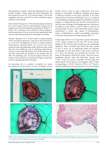
Figure 2: Brief summary of the measurements taken during the different phases of the study performed by the authors’ team. (A) intraoperative
measurement in fresh; (B) measurement in fresh by the pathologist; (C) measurement after fixation with formaldehyde; (D) measurement under microscope
(C) in a patient with oral squamous cell carcinoma - T1N0M0
Plast Aesthet Res || Volume 3 || May 25, 2016 155