
Page 81 - Read Online
P. 81
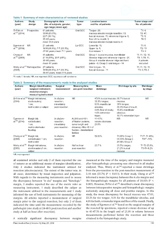
Table 1: Summary of main characteristics of reviewed studies
            Authors   Study      Demographic data        Tumor    Location tumor                  Tumor stage and
            (year)    design     (No. of subjects; gender;   type  and No. of patients             No. of patients
                                 age range; mean age)
           El-Fole et   Prospective  61 patients;      Oral SCC  Tongue: 20;                           T1: 4;
            [23]
           al.  (2014)          39 M (63.9%),                    mucosa alveolar margin mandible: 13;  T2: 47;
                                22 F (36.1%);                    buccal mucosa: 15; retromolar trigone: 6;  T3: 5;
                                35-69 years;                     floor of the mouth: 3;                T4: 5
                                mean 51.6 years                  mucosa alveolar margin maxilla: 4
           Egemen et   NR       21 patients;           Lip SCC   Lower lip: 15;                        T1: 8;
           al.  (2014)          14 M (66.6%), 7 F (33.3%);       Upper lip: 5;                        T2: 10
            [24]
                                47-92 years; mean 71.1 years     Commisure: 1                          T3: 3
           Cheng et   NR        41 patients;           Oral SCC  Group 1: buccal mucosa, mandibular   T1: 11; T2: 16;
            [20]
           al.  (2008)          21 M (51%), 20 F (49%);          Alveolar ridge and retromolar trigone - 21;  T3: 1; T4: 11;
                                35-95 years;                     Group 2: maxilar alveolar ridge and hard   2 patients
                                mean 67 years                    palate - 6; Group 3: oral tongue - 14  excluded
                   [16]
           Mistry et al.  Retrospective 27 patients;   Oral SCC  Oral tongue - 16;                  T1: 11; T2: 11;
           (2005)               18 M (66.6%), 9 F (33.3%);       buccal mucosa - 11                  T3: 3; T4: 2
                                36-61 years; mean 47.6 years
           M: male; F: female; NR: not reported; SCC: squamous cell carcinoma
           Table 2: Summary of the measurements made in the analyzed studies
            Authors   Margin identification;   Surgical   Mearuing time,  Mean        Shrinkage by site  Shrinkage
            (year)    surgical instrument;   measurement  pre-post resection  shrinkage                  by stage
                      resection margin;
                      measuring instrument
           El-Fole et al.  Margin ink/sutures;   In situ/pre-  NR    47.6% buccal mucosa; 66.7% buccal    NR
                   [23]
           (2014)    electocautery;     resection;                   33.3% tongue;   mucosa;
                     1 cm;              inmediately                  9.5% mandibular   35% tongue;
                     metric ruler or caliper  post-resection         alveolus;       33.3% floor of mouth;
                                                                     4.8% floor of mouty;  15.4% mandibular
                                                                     4.8% retromolar   alveolus;
                                                                     trigon          16.7% retromolar trigon
           Egemen et   Margin ink;      In situ/pre-  At 24 h and 48 h   41-47%;     NR                   NR
            [24]
           al.  (2014)  not indicated;  resection;   of fixation (volume,   volume decrease
                     1 cm;              inmediately   tumor length and   21.8%
                     metric ruler/water   post-resection  distance); After 48 h of
                     flooding                        fixation - standardized
                                                     fashion
                   [20]
           Cheng et al.   Margin ink;   In situ/pre-  NR             59.02%          71.90% Group 1;  T1/T -51.48%;
           (2008)    not indicated; 1 cm;   resection                                53.33% Group 2;    T3/T -75%
                     metric ruler                                                    41.14% Group 3
                   [16]
           Mistry et al.   Margin ink/sutures;  In situ/pre-  Half on hour   22.7 %   23.5% tongue;   T1-T2-25.6%;
           (2005)    electrocautery1 cm;  resection  post-resection                  21.2% buccal      T3-T4-9.2%
                     caliper                                                         mucosa
           NR: not reported

           all examined articles and only 2 of them reported the use   measured at the time of the surgery and margins measured
           of sutures as an additional means of margins identification.   after histopathologic processing was observed in all studies
           Only  2 studies  indicated the  instrument  utilized  for   analyzed. Thus, Mistry  et al. [16]  reported a mean shrinkage
           resection (electrocautery). The extent of the tumor was, in   from the pre-resection to the post-resection measurement of
                                                                                                              [20]
           all cases, determined  by  visual inspection and palpation.   3.18 mm (22.7%) (P < 0.011). In their study, Cheng et al.
           With regards to the measuring instruments used to assess   informed a mean discrepancy between the in situ margins and
           the discrepancy between “in situ” margins and “histologic”   the histopathologic margins for all patients of 59.02% (P <
           margin, 3 studies reported the use of the metric ruler as   0.001). However, El-Fol et al.  described a mean discrepancy
                                                                                      [23]
           measuring instrument,  1 study described the caliper  as   between intraoperative margins and histopathologic margins
           the instrument utilized in the measurements and 1 study   exclusively analyzing all close and positive margins. In this
           informed the use of both instruments for measuring of the   study, the mean discrepancy for buccal mucosa was 47.6%,
           margins.  All articles reported a measurement  of surgical   33.3% for the tongue, 9.5% for the mandibular alveolus, and
           margin prior to the surgical resection, but only 2 of them   4.8% for both, retromolar trigon and floor of the mouth. Finally,
                                                                                    [24]
           indicated the time until the measurement recorded by the   the study of Egemen et al.  based on the surgical margins of
           pathologist (one study at 24-48 h postresection and another   the resected lip specimens, reported a mean decrease of up
           study at half an hour after resection).             to 41-47.5% in the length and of 21.8% in volume between
                                                               measurements performed before the resection and those
           A statically significant discrepancy between margins   obtained in the histopatologic study.
           Plast Aesthet Res || Volume 3 || May 25, 2016                                                      153