
Page 16 - Read Online
P. 16
Deldar et al. Plast Aesthet Res 2022;9:13 https://dx.doi.org/10.20517/2347-9264.2021.100 Page 9 of 16
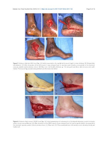
Figure 7. Abductor hallucis (AH) local flap. (A) Initial presentation of a medial ankle wound; depth is down to bone. (B) Wound after
debridement. (C) After dissection of the AH muscle, it was released from its insertion point distally; arrow points to the dominant
vascular pedicle (medial plantar artery). (D) Flap inset. (E) Results at follow-up. The skin graft and donor site incisions were well-
healed. The muscle flap provided adequate bulk over the bony heel defect.
Figure 8. Abductor digiti minimi (ADM) local flap. (A) Initial presentation of osteomyelitis of the lateral calcaneus; purple line marks
where incision was performed. (B) After dissection of the ADM muscle, it was released from its insertion point distally; arrow points to
the dominant vascular pedicle (lateral plantar artery). (C) Flap inset. (D) The wound and donor site incisions were closed primarily and
healed well.