
Page 19 - Read Online
P. 19
Page 12 of 16 Deldar et al. Plast Aesthet Res 2022;9:13 https://dx.doi.org/10.20517/2347-9264.2021.100
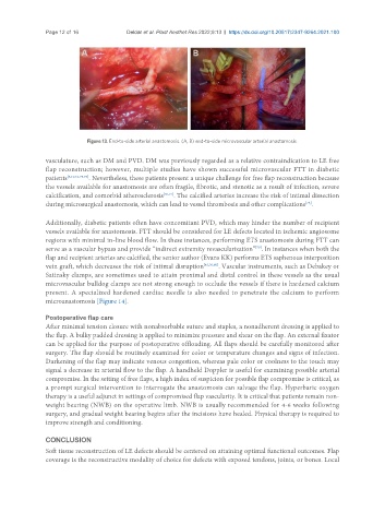
Figure 13. End-to-side arterial anastomosis. (A, B) end-to-side microvascular arterial anastomosis.
vasculature, such as DM and PVD. DM was previously regarded as a relative contraindication to LE free
flap reconstruction; however, multiple studies have shown successful microvascular FTT in diabetic
patients [5,10,63,75,76] . Nevertheless, these patients present a unique challenge for free flap reconstruction because
the vessels available for anastomosis are often fragile, fibrotic, and stenotic as a result of infection, severe
calcification, and comorbid atherosclerosis [50,77] . The calcified arteries increase the risk of intimal dissection
during microsurgical anastomosis, which can lead to vessel thrombosis and other complications .
[78]
Additionally, diabetic patients often have concomitant PVD, which may hinder the number of recipient
vessels available for anastomosis. FTT should be considered for LE defects located in ischemic angiosome
regions with minimal in-line blood flow. In these instances, performing ETS anastomosis during FTT can
serve as a vascular bypass and provide “indirect extremity revascularization” . In instances when both the
[79]
flap and recipient arteries are calcified, the senior author (Evans KK) performs ETS saphenous interposition
vein graft, which decreases the risk of intimal disruption [43,50,80] . Vascular instruments, such as Debakey or
Satinsky clamps, are sometimes used to attain proximal and distal control in these vessels as the usual
microvascular bulldog clamps are not strong enough to occlude the vessels if there is hardened calcium
present. A specialized hardened cardiac needle is also needed to penetrate the calcium to perform
microanastomosis [Figure 14].
Postoperative flap care
After minimal tension closure with nonabsorbable suture and staples, a nonadherent dressing is applied to
the flap. A bulky padded dressing is applied to minimize pressure and shear on the flap. An external fixator
can be applied for the purpose of postoperative offloading. All flaps should be carefully monitored after
surgery. The flap should be routinely examined for color or temperature changes and signs of infection.
Darkening of the flap may indicate venous congestion, whereas pale color or coolness to the touch may
signal a decrease in arterial flow to the flap. A handheld Doppler is useful for examining possible arterial
compromise. In the setting of free flaps, a high index of suspicion for possible flap compromise is critical, as
a prompt surgical intervention to interrogate the anastomosis can salvage the flap. Hyperbaric oxygen
therapy is a useful adjunct in settings of compromised flap vascularity. It is critical that patients remain non-
weight bearing (NWB) on the operative limb. NWB is usually recommended for 4-6 weeks following
surgery, and gradual weight bearing begins after the incisions have healed. Physical therapy is required to
improve strength and conditioning.
CONCLUSION
Soft tissue reconstruction of LE defects should be centered on attaining optimal functional outcomes. Flap
coverage is the reconstructive modality of choice for defects with exposed tendons, joints, or bones. Local