
Page 15 - Read Online
P. 15
Page 8 of 16 Deldar et al. Plast Aesthet Res 2022;9:13 https://dx.doi.org/10.20517/2347-9264.2021.100
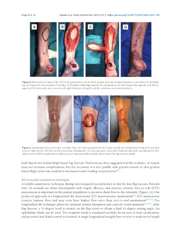
Figure 5. Reversed sural artery flap. (A) Initial presentation of calcaneal wound, flap was designed based on sural artery on posterior
leg. (B) Dissection and elevation of the flap. (C) Rotation of the flap towards the calcaneal wound. (D) Healed flap; superior and inferior
aspects of the donor site were covered with split-thickness skin graft, and the remainder was closed primarily.
Figure 6. Saphenous artery perforator propeller flap. (A) Initial presentation of circular wound over distal anterior leg with exposed
anterior tibial tendon. (B) Wound after excisional debridement. (C) Flap was inset, and a split-thickness skin graft was placed over the
lateral portion of the wound where tendon was not exposed and the medial defect where the flap was harvested.
both lateral and medial thigh-based flap harvest. Furthermore, they suggested that the inclusion of muscle
does not increase complications, but the inclusion of a skin paddle with gracilis muscle or skin grafted
lateral thigh donor site resulted in increased wound healing complications .
[70]
Microvascular anastomotic techniques
A reliable anastomotic technique during microsurgical reconstruction is vital for free flap success. Patients
with LE wounds are often vasculopaths with fragile, fibrotic, and stenotic arteries. End-to-side (ETS)
anastomosis is important in this patient population to preserve distal flow to the extremity [Figure 13]. Our
preferred approach is a longitudinal slit arteriotomy ETS microvascular anastomosis . ETS anastomosis
[43]
ensures laminar flow and may even have higher flow rates than end-to-end anastomosis [43,71,72] . The
longitudinal slit technique allows for minimal intimal disruption and controls vessel mismatch [43,71,73] . After
flap harvest, a 70-degree bevel is created on the flap artery to obtain a final 20-degree resting angle. An
ophthalmic blade can be used. The recipient vessel is examined carefully for an area of least calcification,
and proximal and distal control is obtained. A single longitudinal straight-line incision is made to the length