
Page 18 - Read Online
P. 18
Deldar et al. Plast Aesthet Res 2022;9:13 https://dx.doi.org/10.20517/2347-9264.2021.100 Page 11 of 16
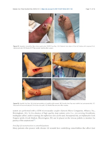
Figure 11. Superior circumflex iliac artery perforator (SCIP) free flap. (A) Diabetic foot ulcer of the left hallux with exposed first
metatarsal joint. (B) Healed SCIP flap several months after surgery.
Figure 12. Gracilis free flap. (A) Initial presentation of medial ankle wound. (B) Gracilis free flap was marked out preoperatively. (C)
Flap inset with an overlying split-thickness skin graft. (D) Healed flap one year after surgery.
system are performed with a GEM microvascular coupler (Synovis Micro Companies, Alliance, Inc.,
Birmingham, AL). In the absence of high-quality deep-system veins (i.e., pre-existing thrombosis,
inadequate caliber, and/or scarring), the saphenous vein can be used. Intraoperatively, an implantable Cook
Doppler probe (Cook Medical, Bloomington, IN) can be placed on the venous pedicle to monitor the
patency of the anastomosis .
[74]
Free flap LE reconstruction in comorbid patients
Many patients who present with chronic LE wounds have underlying comorbidities that affect their