
Page 13 - Read Online
P. 13
Page 6 of 16 Deldar et al. Plast Aesthet Res 2022;9:13 https://dx.doi.org/10.20517/2347-9264.2021.100
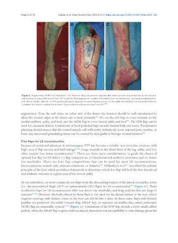
Figure 2. Angiosomes of the foot and ankle. (A) Anterior tibial angiosome: supplies the anterior ankle and continues as the dorsalis
pedis artery to supply the dorsal foot. (B) Posterior tibial angiosome: supplies the plantar foot via 3 branches: calcaneal, medial plantar,
and lateral plantar arteries. (C) Peroneal angiosome supplies the anterolateral portion of the ankle and hindfoot via 2 branches: lateral
calcaneal and anterior perforator arteries. Figure adapted with permission from Ref. [49] .
angiosomes. Thus, the soft tissue on either side of the donor site incision should be well-vascularized to
allow the wound edges at the donor site to heal primarily . We use the AH flap to cover wounds on the
[55]
[55]
medial midfoot, ankle, and heel, and the ADM flap to cover lateral ankle and foot . The FDB flap can be
used for calcaneal defects. Limitations of local pedicled flaps include limited bulk and reach. Preoperative
planning should ensure that the rotated muscle will sufficiently definitively cover exposed joint, tendon, or
bone; any uncovered granulating tissue can be covered by skin grafts or biologic wound matrices .
[55]
Free flaps for LE reconstruction
Because of continued advances in microsurgery, FTT has become a reliable reconstructive solution, with
high rates of flap success and limb salvage [63,64] . Large wounds in the distal third of the leg, ankle, and foot
[65]
often require free tissue reconstruction . There are three main considerations to guide the choice of
optimal free flap for LE defect: (1) flap composition; (2) functional and aesthetic outcomes; and (3) donor
site morbidity. There are four flap compositions that can be used for most LE reconstructions:
fasciocutaneous, muscle only, musculocutaneous, or chimeric . Hollenbeck et al. described the subunit
[66]
[67]
principle of the foot, which provides a framework to determine which free flap will lend the best functional
and aesthetic outcome in a given area of the foot or ankle.
At our institution, we most commonly use flaps from the descending branch of the lateral circumflex artery
[i.e., the anterolateral thigh (ALT) or vastus lateralis (VL) flaps] for LE reconstruction [Figure 10]. These
[50]
workhorse flaps for LE reconstruction offer low donor site morbidity and long pedicles that are large in
diameter [50,68] . However, the bulk offered by these flaps is not ideal for the dorsal surface of the foot, which
requires coverage with thinner tissue so the foot can still fit into a shoe. In these cases, flaps with thinner
paddles are preferred: the radial forearm flap, MSAP flap, or superior circumflex iliac artery perforator
(SCIP) flap are reasonable options [50,59] [Figure 11]. Limitations of the SCIP flap include a short, small caliber
pedicle, while the MSAP flap requires tedious muscle dissection and susceptibility to vein damage given the