
Page 11 - Read Online
P. 11
Page 4 of 16 Deldar et al. Plast Aesthet Res 2022;9:13 https://dx.doi.org/10.20517/2347-9264.2021.100
Table 1. Thrombophilia screening panel
Basic labs
• CBC
• PT/INR
• PTT
Hypercoagulable tests
• Factor V Leiden G1691A genotype
• Prothrombin G20210A genotype
• Homocysteine level
• Factor VIII level
• Antiphospholipid antibody testing
• Antithrombin III activity
• Protein C activity
• Protein S activity
• MTHFR polymorphisms (A1298C and C677T)
• PAI-1 4G/5G QST
CBC: Complete blood count; PT/INR: prothrombin time/international normalized ratio; PTT: partial thromboplastin time; MTHFR:
methylenetetrahydrofolate reductase; PAI-1: plasminogen activator inhibitor 1; QST: quantitative sensory testing.
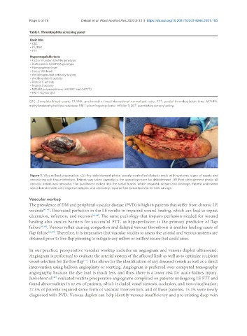
Figure 1. Wound bed preparation. (A) Pre-debridement photo: poorly-controlled diabetic male with systemic signs of sepsis and
necrotizing soft tissue infection. Patient was taken urgently to the operating room for debridement. (B) Post-debridement photo: all
necrotic debris was removed. The purulence tracked into the tarsal tunnel, which required incision and drainage. Patient underwent
serial debridements until negative cultures, and ultimately required free tissue transfer for limb salvage.
Vascular workup
The prevalence of DM and peripheral vascular disease (PVD) is high in patients that suffer from chronic LE
wounds [41-43] . Decreased perfusion in the LE results in impaired wound healing, which can lead to repeat
ulceration, infection, and necrosis [41,44] . The same pathology that impairs perfusion needed for wound
healing also creates barriers for successful FTT, as hypoperfusion is the primary predictor of flap
failure [41,45] . Venous reflux causing congestion and delayed venous thrombosis is another leading cause of
flap failure [46,47] . Therefore, it is imperative that vascular studies to assess the arterial and venous systems are
obtained prior to free flap planning to mitigate any inflow or outflow issues that could arise.
In our practice, preoperative vascular workup includes an angiogram and venous duplex ultrasound.
Angiogram is performed to evaluate the arterial system of the affected limb as well as to optimize recipient
vessel selection for the free flap . This allows for the identification of any diseased vessels as well as a direct
[41]
intervention using balloon angioplasty or stenting. Angiogram is preferred over computed tomography
angiography because the dye load is much less, and thus, there is a lower risk for acute kidney injury.
Janhoferet al. evaluated routine preoperative angiograms completed on patients undergoing LE FTT and
[41]
found abnormalities in 67.8% of patients, which included vessel stenosis, occlusion, and non-visualization;
27.5% of patients required some form of vascular intervention, and of these patients, 15.3% were newly
diagnosed with PVD. Venous duplex can help identify venous insufficiency and pre-existing deep vein