
Page 16 - Read Online
P. 16
Page 2 of 11 Suami et al. Plast Aesthet Res 2019;6:33 I http://dx.doi.org/10.20517/2347-9264.2019.46
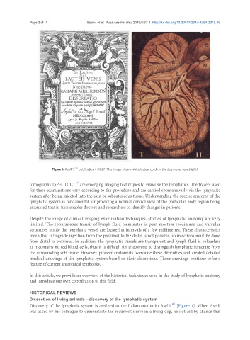
Figure 1. Aselli’s [10] publication in 1627. This image shows white lacteal cords in the dog mesentery (right)
[9]
tomography (SPECT)/CT are emerging imaging techniques to visualise the lymphatics. The tracers used
for these examinations vary according to the procedure and are carried spontaneously via the lymphatic
system after being injected into the skin or subcutaneous tissue. Understanding the precise anatomy of the
lymphatic system is fundamental for providing a normal control view of the particular body region being
examined that in turn enables doctors and researchers to identify changes in patients.
Despite the range of clinical imaging examination techniques, studies of lymphatic anatomy are very
limited. The spontaneous transit of lymph fluid terminates in post-mortem specimens and valvular
structures inside the lymphatic vessel are located at intervals of a few millimetres. These characteristics
mean that retrograde injection from the proximal to the distal is not possible, so injections must be done
from distal to proximal. In addition, the lymphatic vessels are transparent and lymph fluid is colourless
as it contains no red blood cells, thus it is difficult for anatomists to distinguish lymphatic structure from
the surrounding soft tissue. However, pioneer anatomists overcame these difficulties and created detailed
medical drawings of the lymphatic system based on their dissections. These drawings continue to be a
feature of current anatomical textbooks.
In this article, we provide an overview of the historical techniques used in the study of lymphatic anatomy
and introduce our own contribution to this field.
HISTORICAL REVIEWS
Dissection of living animals - discovery of the lymphatic system
[10]
Discovery of the lymphatic system is credited to the Italian anatomist Aselli [Figure 1]. When Aselli
was asked by his colleague to demonstrate the recurrent nerve in a living dog, he noticed by chance that