
Page 21 - Read Online
P. 21
Suami et al. Plast Aesthet Res 2019;6:33 I http://dx.doi.org/10.20517/2347-9264.2019.46 Page 7 of 11
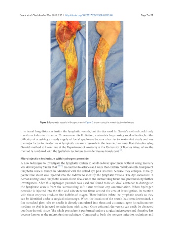
Figure 6. Lymphatic vessels in the specimen in Figure 5 shown using the microinjection technique
it to travel long distances inside the lymphatic vessels, but the dye used in Gerota’s method could only
travel much shorter distances. To overcome this limitation, anatomists began using smaller bodies, but the
difficulty of acquiring a steady supply of foetal specimens became a barrier to anatomical study and was
the major factor in the decline of lymphatic anatomy research in the twentieth century. Foetal studies using
Gerota’s method still continue at the Department of Anatomy at the University of Buenos Aires, where the
[25]
method is combined with the Spalteholz technique to render tissues translucent .
Microinjection technique with hydrogen peroxide
A new technique to investigate the lymphatic system in adult cadaver specimens without using mercury
was developed by Suami et al. [26,27] . In contrast to arteries and veins that contain red blood cells, transparent
lymphatic vessels cannot be identified with the naked eye post-mortem because they collapse. Initially,
patent blue violet was injected into the cadaver to identify the lymphatic vessels. The dye succeeded in
demonstrating some lymphatic vessels, but it also stained the surrounding tissue and prevented any further
investigation. After this, hydrogen peroxide was used and found to be an ideal substance to distinguish
the lymphatic vessels from the surrounding soft tissue without any contamination. When hydrogen
peroxide is injected into the skin and subcutaneous tissue around the area of investigation, its reaction
with tissue enzymes produces fine bubbles of oxygen. These bubbles inflate the lymphatic vessels so they
can be identified under a surgical microscope. When the location of the vessels has been determined, a
fine stretched glass tube or needle is directly cannulated into them and a contrast agent (a radiocontrast
medium or dye) is injected to stain them with colour. Once coloured, the vessels can easily be dissected
out from the soft tissue. The whole procedure is performed under a surgical microscope and therefore has
become known as the microinjection technique. Compared to both the mercury injection technique and