
Page 17 - Read Online
P. 17
Suami et al. Plast Aesthet Res 2019;6:33 I http://dx.doi.org/10.20517/2347-9264.2019.46 Page 3 of 11
[13]
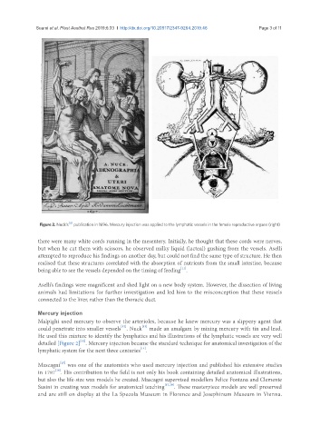
Figure 2. Nuck’s publication in 1696. Mercury injection was applied to the lymphatic vessels in the female reproductive organs (right)
there were many white cords running in the mesentery. Initially, he thought that these cords were nerves,
but when he cut them with scissors, he observed milky liquid (lacteal) gushing from the vessels. Aselli
attempted to reproduce his findings on another day, but could not find the same type of structure. He then
realised that these structures correlated with the absorption of nutrients from the small intestine, because
[11]
being able to see the vessels depended on the timing of feeding .
Aselli’s findings were magnificent and shed light on a new body system. However, the dissection of living
animals had limitations for further investigation and led him to the misconception that these vessels
connected to the liver, rather than the thoracic duct.
Mercury injection
Malpighi used mercury to observe the arterioles, because he knew mercury was a slippery agent that
[13]
[12]
could penetrate into smaller vessels . Nuck made an amalgam by mixing mercury with tin and lead.
He used this mixture to identify the lymphatics and his illustrations of the lymphatic vessels are very well
[13]
detailed [Figure 2] . Mercury injection became the standard technique for anatomical investigation of the
[14]
lymphatic system for the next three centuries .
[15]
Mascagni was one of the anatomists who used mercury injection and published his extensive studies
[16]
in 1787 . His contribution to the field is not only his book containing detailed anatomical illustrations,
but also the life-size wax models he created. Mascagni supervised modellers Felice Fontana and Clemente
Susini in creating wax models for anatomical teaching [17,18] . These masterpiece models are well preserved
and are still on display at the La Specola Museum in Florence and Josephinum Museum in Vienna.