
Page 20 - Read Online
P. 20
Page 6 of 11 Suami et al. Plast Aesthet Res 2019;6:33 I http://dx.doi.org/10.20517/2347-9264.2019.46
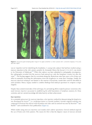
Figure 5. Indocyanine green lymphography image in the upper extremity in a fresh cadaver (left). Lymphatic mapping in the same
specimen (right)
was an imperfect tool for identifying the lymphatics. A young male cadaver that had been studied using a
mercury injection in the 18th century, possibly by Alexander Monro the Second (1733-1817), was stored
[20]
at the University of Edinburgh . When this cadaver was later submitted to radiographic investigation,
the radiographs revealed that the mercury had entered not only the lymphatic vessels, but also the
[21]
veins . This finding suggests that the anatomists doing the illustrations must have spent a lot of time and
effort in manually extracting the information about the lymphatics to create their drawings. Finally, the
mercury injection technique was limited in the number of lymphatic vessels that could be identified in
each specimen. Hence, Sappey needed to combine findings from multiple specimens to compose a single
diagram.
Despite these several downsides of the technique, the painstaking efforts made by pioneer anatomists who
used mercury injection succeeded in establishing the solid foundation of lymphatic anatomy for three
centuries, and our current knowledge still relies heavily on their achievement.
Dye injection
As a potential replacement for mercury injection, a dye injection method for demonstrating the lymphatics
[22]
was developed by Gerota in a technique known as Gerota’s method. Gerota’s original medium was
[23]
composed of Prussian blue diluted with turpentine and ether and his method was used by Rouvière and
Bartels and contributed to their publications [Figure 4].
[24]
While studies using mercury injection used mainly adult cadaver specimens, Gerota’s method required
the use of foetal and child cadavers. The reason for this is that the slippery nature of mercury allowed