
Page 23 - Read Online
P. 23
Suami et al. Plast Aesthet Res 2019;6:33 I http://dx.doi.org/10.20517/2347-9264.2019.46 Page 9 of 11
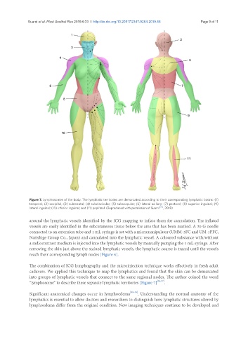
Figure 7. Lymphosomes of the body. The lymphatic territories are demarcated according to their corresponding lymphatic basins: (1)
temporal; (2) occipital; (3) submental; (4) subclavicular; (5) subscapular; (6) lateral axillary; (7) pectoral; (8) superior inguinal; (9)
lateral inguinal; (10) inferior inguinal; and (11) popliteal. (Reproduced with permission of Suami [37] , 2018)
around the lymphatic vessels identified by the ICG mapping to inflate them for cannulation. The inflated
vessels are easily identified in the subcutaneous tissue below the area that has been marked. A 30 G needle
connected to an extension tube and 1 mL syringe is set with a micromanipulator (UMM-3FC and UM-1PFC,
Narishige Group Co., Japan) and cannulated into the lymphatic vessel. A coloured substance with/without
a radiocontrast medium is injected into the lymphatic vessels by manually pumping the 1 mL syringe. After
removing the skin just above the stained lymphatic vessels, the lymphatic course is traced until the vessels
reach their corresponding lymph nodes [Figure 6].
The combination of ICG lymphography and the microinjection technique works effectively in fresh adult
cadavers. We applied this technique to map the lymphatics and found that the skin can be demarcated
into groups of lymphatic vessels that connect to the same regional nodes. The author coined the word
“lymphosome” to describe these separate lymphatic territories [Figure 7] [36,37] .
Significant anatomical changes occur in lymphoedema [38-40] . Understanding the normal anatomy of the
lymphatics is essential to allow doctors and researchers to distinguish how lymphatic structures altered by
lymphoedema differ from the original condition. New imaging techniques continue to be developed and