
Page 19 - Read Online
P. 19
Suami et al. Plast Aesthet Res 2019;6:33 I http://dx.doi.org/10.20517/2347-9264.2019.46 Page 5 of 11
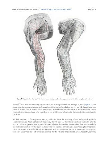
Figure 4. Illustration from Bartels’ [24] book showing lymphatic vessels in the upper extremity identified using Gerota’s method
[19]
Sappey also used the mercury injection technique and published his findings in 1874 [Figure 3]. His
book provided a comprehensive understanding of the human lymphatics, but his superb illustrations were
more of artistic than scientific value. Sappey was probably the first anatomist to understand the idea of
lymphatic territories defined by a watershed at the midline and a horizontal line crossing the umbilicus in
the torso.
To date, anatomical findings with mercury injection were the mainstay of our understanding of the
lymphatic system. Anatomists injected mercury directly into the lymphatic vessels or indirectly into the
skin in cadaveric specimens using stretched glass tubes or fine needles. The excellent illustrations made by
the early anatomists led to the belief that mercury was an ideal material for demonstrating the lymphatics,
but it has several downsides. Firstly, mercury is a toxic substance and its use in anatomical investigation
was discontinued in the early twentieth century due to concerns about health issues. Secondly, mercury