


Page 36 - Read Online
P. 36
Page 10 of 18             Rodriguez. Plast Aesthet Res 2023;10:67  https://dx.doi.org/10.20517/2347-9264.2023.73


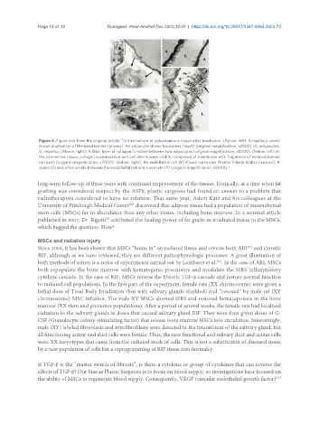
                Figure 6. Figure text from the original article: “Ultrastructure of subcutaneous tissue after irradiation. (Above, left): A capillary vessel
                shows duplication of the basal lamina (arrows). An adipocyte shows lysosomes (inset) (original magnification, x2500) (A, adipocytes;
                V, vessels); (Above, right): A thick layer of collagen is visible between two adipocytes (original magnification, x5000). (Below, left): In
                the connective tissue, collagen accumulation and cell debris were visible, composed of membrane with fragments of external lamina
                (arrows) (original magnification, x7000); (Below, right): An endothelial cell (E) shows numerous Weibel-Palade bodies (arrows); A
                space (S) was often visible between the endothelial cell and a pericyte (P) (original magnification, x8000).”

               long-term follow-up of three years with continued improvement of the tissues. Ironically, at a time when fat
               grafting was considered suspect by the ASPS, plastic surgeons had found an answer to a problem that
               radiotherapists considered to have no solution. That same year, Adam Katz and his colleagues at the
               University of Pittsburgh Medical Center  discovered that adipose tissue had a population of mesenchymal
                                                 [24]
               stem cells (MSCs) far in abundance than any other tissue, including bone marrow. In a seminal article
               published in 2007, Dr. Rigotti  attributed the healing power of fat grafts in irradiated tissue to the MSCs,
                                         [2]
               which begged the question- How?


               MSCs and radiation injury
               Since 2003, it has been shown that MSCs “home in” on radiated tissue and reverse both ARI  and chronic
                                                                                             [25]
               RIF, although as we have reviewed, they are different pathophysiologic processes. A great illustration of
               both methods of action is a series of experiments carried out by Lombaert et al. . In the case of ARI, MSCs
                                                                                  [26]
               both repopulate the bone marrow with hematogenic precursors and modulate the SIRS inflammatory
               cytokine cascade. In the case of RIF, MSCs reverse the fibrotic TGF-β cascade and restore normal function
               to radiated cell populations. In the first part of the experiment, female rats (XX chromosome) were given a
               lethal dose of Total Body Irradiation (but with salivary glands shielded) and “rescued” by male rat (XY
               chromosome) MSC infusion. The male XY MSCs aborted SIRS and restored hematopoiesis in the bone
               marrow (XX stem and precursor populations). After a period of several weeks, the female rats had localized
               radiation to the salivary glands in doses that caused salivary gland RIF. They were then given doses of G-
               CSF (Granulocyte colony-stimulating factor) that release bone marrow MSCs into circulation. Interestingly,
               male (XY) labeled fibroblasts and myofibroblasts were detected in the interstitium of the salivary gland, but
               all functioning acinar and duct cells were female. Thus, the new functional and salivary duct and acinar cells
               were XX karyotypes that came from the radiated stock of cells. This is not a substitution of diseased tissue
               by a new population of cells but a reprogramming of RIF tissue into normalcy.


               If TGF-β is the “master switch of fibrosis”, is there a cytokine or group of cytokines that can reverse the
               effects of TGF-β? Our bias as Plastic Surgeons is to focus on blood supply, so investigations have focused on
                                                                                                        [27]
               the ability of MSCs to regenerate blood supply. Consequently, VEGF (vascular endothelial growth factor)

