


Page 38 - Read Online
P. 38
Page 12 of 18             Rodriguez. Plast Aesthet Res 2023;10:67  https://dx.doi.org/10.20517/2347-9264.2023.73


                            [28]
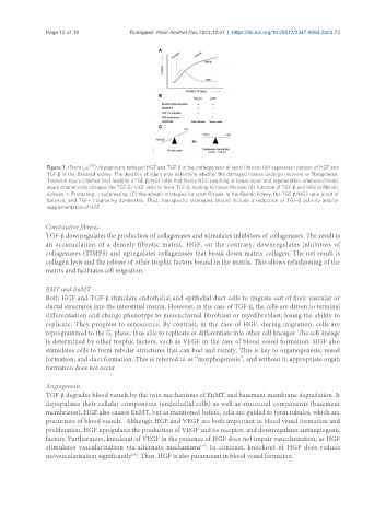
               Figure 7. (From Liu  ) Antagonism between HGF and TGF-β in the pathogenesis of renal fibrosis. (A) expression pattern of HGF and
               TGF-β in the diseased kidney. The duration of injury may determine whether the damaged tissues undergo recovery or fibrogenesis.
               Transient injury (dashed line) leads to a TGF-β/HGF ratio that favors HGF, resulting in tissue repair and regeneration, whereas chronic
               injury dramatically changes the TGF-β/ HGF ratio to favor TGF-β, leading to tissue fibrosis; (B) function of TGF-β and HGF in fibrotic
               kidneys. +: Promoting; -: suppressing; (C) therapeutic strategies for renal fibrosis. In the fibrotic kidney, the TGF-β/HGF ratio is out of
               balance, and TGF- 1 signaling dominates. Thus, therapeutic strategies should include a reduction of TGF-β activity and/or
               supplementation of HGF.


               Constitutive fibrosis
               TGF-β downregulates the production of collagenases and stimulates inhibitors of collagenases. The result is
               an accumulation of a densely fibrotic matrix. HGF, on the contrary, downregulates inhibitors of
               collagenases (TIMPS) and upregulates collagenases that break down matrix collagen. The net result is
               collagen lysis and the release of other trophic factors bound in the matrix. This allows refashioning of the
               matrix and facilitates cell migration.


               EMT and EnMT
               Both HGF and TGF-β stimulate endothelial and epithelial duct cells to migrate out of their vascular or
               ductal structures into the interstitial matrix. However, in the case of TGF-β, the cells are driven to terminal
               differentiation and change phenotype to mesenchymal fibroblast or myofibroblast, losing the ability to
               replicate. They progress to senescence. By contrast, in the case of HGF, during migration, cells are
               reprogrammed to the G  phase, thus able to replicate or differentiate into other cell lineages. The cell lineage
                                   0
               is determined by other trophic factors, such as VEGF in the case of blood vessel formation. HGF also
               stimulates cells to form tubular structures that can bud and ramify. This is key to organogenesis, vessel
               formation, and duct formation. This is referred to as “morphogenesis”, and without it, appropriate organ
               formation does not occur.


               Angiogenesis
               TGF-β degrades blood vessels by the twin mechanisms of EnMT and basement membrane degradation. It
               depopulates their cellular components (endothelial cells) as well as structural components (basement
               membranes). HGF also causes EnMT, but as mentioned before, cells are guided to form tubules, which are
               precursors of blood vessels.  Although HGF and VEGF are both important to blood vessel formation and
               proliferation, HGF upregulates the production of VEGF and its receptor, and downregulates antiangiogenic
               factors. Furthermore, knockout of VEGF in the presence of HGF does not impair vascularization, as HGF
                                                               [34]
               stimulates vascularization via alternate mechanisms . In contrast, knockout of HGF does reduce
               neovascularization significantly . Thus, HGF is also paramount in blood vessel formation.
                                          [35]

