


Page 34 - Read Online
P. 34
Page 8 of 18              Rodriguez. Plast Aesthet Res 2023;10:67  https://dx.doi.org/10.20517/2347-9264.2023.73


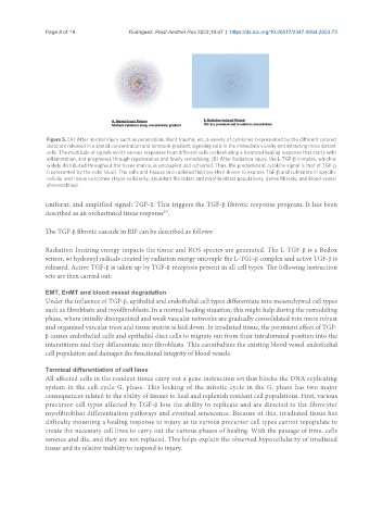
                Figure 5. (A) After normal injury such as penetration, blunt trauma,  etc., a variety of cytokines (represented by the different colored
                dots) are released in a spatial concentration and temporal gradient, signaling cells in the immediate vicinity and attracting more distant
                cells. The multitude of signals elicits various responses from different cells orchestrating a balanced healing response that starts with
                inflammation, and progresses through regeneration and finally remodeling; (B) After Radiation injury, the L-TGF-β complex, which is
                widely distributed throughout the tissue matrix, is uncoupled and activated. Thus, the predominant cytokine signal is that of TGF-β
                (represented by the color blue). The cells and tissues in a radiated field are then driven to express TGF-β and culminate in specific
                cellular and tissue outcomes (Hypo cellularity, abundant fibroblast and myofibroblast populations, dense fibrosis, and blood vessel
                abnormalities).


               uniform, and amplified signal: TGF-β. This triggers the TGF-β fibrotic response program. It has been
               described as an orchestrated tissue response .
                                                    [5]

               The TGF-β fibrotic cascade in RIF can be described as follows:


               Radiation Ionizing energy impacts the tissue and ROS species are generated. The L-TGF-β is a Redox
               sensor, so hydroxyl radicals created by radiation energy uncouple the L-TGF-β complex and active TGF-β is
               released. Active TGF-β is taken up by TGF-β receptors present in all cell types. The following instruction
               sets are then carried out:

               EMT, EnMT and blood vessel degradation
               Under the influence of TGF-β, epithelial and endothelial cell types differentiate into mesenchymal cell types
               such as fibroblasts and myofibroblasts. In a normal healing situation, this might help during the remodeling
               phase, where initially disorganized and weak vascular networks are gradually consolidated into more robust
               and organized vascular trees and tissue matrix is laid down. In irradiated tissue, the persistent effect of TGF-
               β causes endothelial cells and epithelial duct cells to migrate out from their intraluminal position into the
               interstitium and they differentiate into fibroblasts. This cannibalizes the existing blood vessel endothelial
               cell population and damages the functional integrity of blood vessels.

               Terminal differentiation of cell lines
               All affected cells in the resident tissue carry out a gene instruction set that blocks the DNA replicating
               system in the cell cycle G  phase. This locking of the mitotic cycle in the G  phase has two major
                                       1
                                                                                     1
               consequences related to the ability of tissues to heal and replenish resident cell populations. First, various
               precursor cell types affected by TGF-β lose the ability to replicate and are directed to the fibrocyte/
               myofibroblast differentiation pathways and eventual senescence. Because of this, irradiated tissue has
               difficulty mounting a healing response to injury as its various precursor cell types cannot repopulate to
               create the necessary cell lines to carry out the various phases of healing. With the passage of time, cells
               senesce and die, and they are not replaced. This helps explain the observed hypocellularity of irradiated
               tissue and its relative inability to respond to injury.

