


Page 31 - Read Online
P. 31
Rodriguez. Plast Aesthet Res 2023;10:67  https://dx.doi.org/10.20517/2347-9264.2023.73  Page 5 of 18


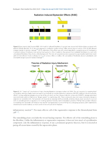
                Figure 2. Experiments that illustrate RIBE. (A) A well of cultured fibroblasts (in green) was covered with Mylar dishes equipped with
                shutters (black) that left 1% of the cells exposed to irradiation (yellow strips). After several days of culture, 30% of cells showed
                changes typical of radiation  damage [10] ; (B) To determine if this effect was cell contact-dependent, a separate group of investigators
                irradiated cultured fibroblasts (red vertical hash pattern). After irradiation, they placed the irradiated fibroblasts in a well with
                unirradiated fibroblasts (blue divots, white background). The two cell preparations were separated by a permeable mesh (black mesh
                pattern) that allowed media exchange but no cell contact. After co-culture, the unirradiated fibroblasts showed cytoplasmic and
                                               [11]
                chromatid changes typical of radiation damage .


                Figure 3. (A) “Target cell” mechanism of injury. Ionizing Radiation damages nuclear cell DNA. The cell responds by repairing itself
                successfully, whereas a faulty repair can result in survival with an unstable genome or apoptosis; (B) RIBE (radiation induced bystander
                effect)- Ionizing radiation damages any portion of the cell which can then release molecular compounds (DAMPS- damage associated
                macromolecular patterns) that act as “danger signals” to neighboring cells. These compounds may even cause chromatin changes in
                adjacent non-irradiated cells. The bystander cell responds by repairing itself successfully, whereas a faulty repair can result in survival
                with an unstable genome or apoptosis; (C) Ionizing radiation liberates matrix-embedded cytokines (TGF-β) that can then influence
                surrounding cells. Bystander cell behavior may then be “reprogrammed” by active TGF-β into undergoing apoptosis, EMT (epithelial to
                mesenchymal transformation), or mitotic arrest of the cell cycle at the G1 phase.


               inflammatory reaction . The main effector cell of the regenerative response is the Mesenchymal Stem
                                   [14]
               cell [15-17] .

               The remodeling phase concludes the wound healing response. The effector cell of the remodeling phase is
               the fibroblast. Unlike the inflammatory or regenerative responses, it does not have much of a proliferative
               component. Like the inflammatory response, it has a prominent apoptotic function, but it is directed at
               paring off excess tissue created by the regenerative phase.

