


Page 29 - Read Online
P. 29
Rodriguez. Plast Aesthet Res 2023;10:67  https://dx.doi.org/10.20517/2347-9264.2023.73  Page 3 of 18


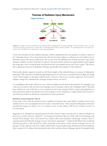
                Figure 1. The target cell theory holds that when radiation hits an individual cell, it sustains damage. The cell attempts repair, or it may
                proceed to apoptosis. Sometimes it survives, but with damaged DNA and an unstable genome. The unstable genome may express as a
                cancer cell, a “crippled” no-functional cell, or may undergo apoptosis.

               Under this rationale, the late radiation damage could be explained as the late sequelae of radiation injury on
               the “bystander tissue”. Since those bystander cells had also been subject to radiation (at a sub-lethal dose for
               bystander tissue), the tissues could repair and recover from the sublethal dose, but the eventual scope of the
               damage would be revealed at the time of mitosis. This post-mitotic attrition in radiated fields would explain
               the progressive loss of normal tissue and function seen in interstitial tissues and blood vessels. This theory
                                                                                           [7]
               led to significant advances in Radiation Therapy, specifically, fractionation of dose delivery .
               Historically, plastic surgeons focused on the blood supply of the radiated tissue as a means of helping the
               tissue heal. This rationale is intuitively appealing because of our focus on vascularized tissue flaps providing
               “fresh” blood supply to damaged radiated tissue. However, this is not a curative approach, but instead a
               palliative designed to substitute the damaged radiated tissue, not heal it.


               As compelling as the target cell theory was, it did not explain several clinical observations. For example, how
               could one account for the severity of late damage seen in bystander tissues (the “bystander effect”) that had
               been radiated at a sub-lethal dose, or for variations in late tissue damage (both in target cell populations as
               well as bystander tissues) not predicted by dose/response curves? Advances in single-particle microbeams to
               cell cultures provided an alternative explanation for these questions .
                                                                        [8]

               Radiation injury bystander effects
               In the early 1990s, with the advent of micro radiation techniques that could deliver radiation doses at the
               cellular level in vitro, investigators focused on the “bystander tissue” effects made startling discoveries that
               upended the target cell theory. For an excellent review on the subject, KM Prise’s excellent review  is
                                                                                                      [9]
               recommended.


               Nagasawa et al. designed an experimental model where barrier strips were placed in a cultured cell plate so
                                               [10]
               that only 1% of the cells were radiated . An unexpected result was that 30% of the cells showed damage and
               nuclear DNA changes. Thus, other factors were at play rather than individual cell damage. Another series of
                                                                         [11]
               experiments showed that the damage was not cell contact-dependent . Cultured fibroblasts were irradiated
               and then placed in a well divided by a filter mesh that would allow culture media exchange but not cell
               passage. On the opposite side were cultured fibroblasts of normal phenotype that had not been irradiated.
               After a period of co-culture, the unirradiated side of the well also showed damage typical of irradiated cells,

