


Page 32 - Read Online
P. 32
Page 6 of 18              Rodriguez. Plast Aesthet Res 2023;10:67  https://dx.doi.org/10.20517/2347-9264.2023.73


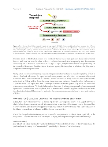
                Figure 4. From the top: Step 1 When tissue is injured, damage signals (DAMPS, Extravasated blood, etc.) are released. Step 2 These
                damage signals are picked up by “sentinel cells”, such as macrophages or mast cells. These cells initiate a cascade of events.  Step 3
                They can amplify the signal by secreting more alarm cytokines and chemoattractants. Step 4 The result is the recruitment of other
                immune effector cells. These can be granulocytes, Lymphocytes, dendritic cells, etc. These immune effector cells can (E) send negative
                or positive feedback signals. Alternately, they can trigger other response pathways and (F) reprogram Affected Tissue in different ways.


               The main point of this brief discussion is to point out that the three main pathways share some common
               features with one but not the other pathway, and the three are linked temporally. But this complex
               relationship can be disrupted by an excess of one type of signal, or by the inability of a cell type to carry out
               its prescribed function. Another factor that can upset this interplay is whether the disorder is
               compartmentalized or generalized.


               Finally, when one of these tissue response patterns goes out of control due to excessive signaling, or lack of
               effective feedback inhibition, the signal amplification process overrides other homeostatic checks and
                      [18]
               balances . These events are known as “cytokine storms” or cascades. Many advanced disease states can be
               understood as falling within these abnormal tissue response patterns. Examples of an inflammatory
               “Cytokine storm” are sepsis and ARS. Although caused by different agents, they share the same final
               common response pathway- “Systemic Inflammatory Response Syndrome”. An example of an unrestrained
               regenerative cascade would be a neoplasm, and an unrestrained remodeling phase can become a fibrotic
               state. Radiation-Induced fibrosis can be understood as one such cascade, precipitated by an overabundance
               of TGF-β.


               HOW THE TGF-β CASCADE CREATES THE TISSUE EFFECTS SEEN IN RIF
               In RIF, the delayed tissue response is not as dependent on dosage and can be seen in patients where
               relatively low doses were administered. It is characterized by persistent fibrosis and varying degrees of loss
               of tissue function. The microscopic appearance typically consists of hypocellularity with a predominance of
               fibroblasts, an abundance of collagen, and blood vessel abnormalities.


               Why is the delayed radiation injury pattern so specific, irrespective of irradiated tissue type? Why is the
               delayed tissue response different than other types of injury, such as penetrating trauma or blast injury?


               The role of TGF-β
                                                              [19]
               TGF-β has been called “the master regulator of fibrosis” . Several characteristics of this cytokine make it a
               good candidate for acting as a “master switch”, specifically in the case of radiation injury.

